Wrist Joint Replacement (Wrist Arthroplasty) OrthoInfo AAOS
Lunate is keystone. The wrist is a unique joint interposed between the distal aspect of the forearm and the proximal aspect of the hand. All three regions have common or shared elements, which integrate form and function to maximize the mechanical effectiveness of the upper extremity. The wrist enables the hand to be placed in an infinite.

Anatomy Of The Wrist Joint
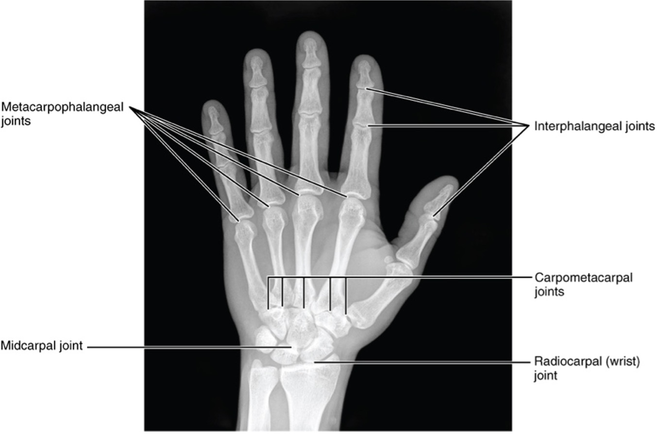
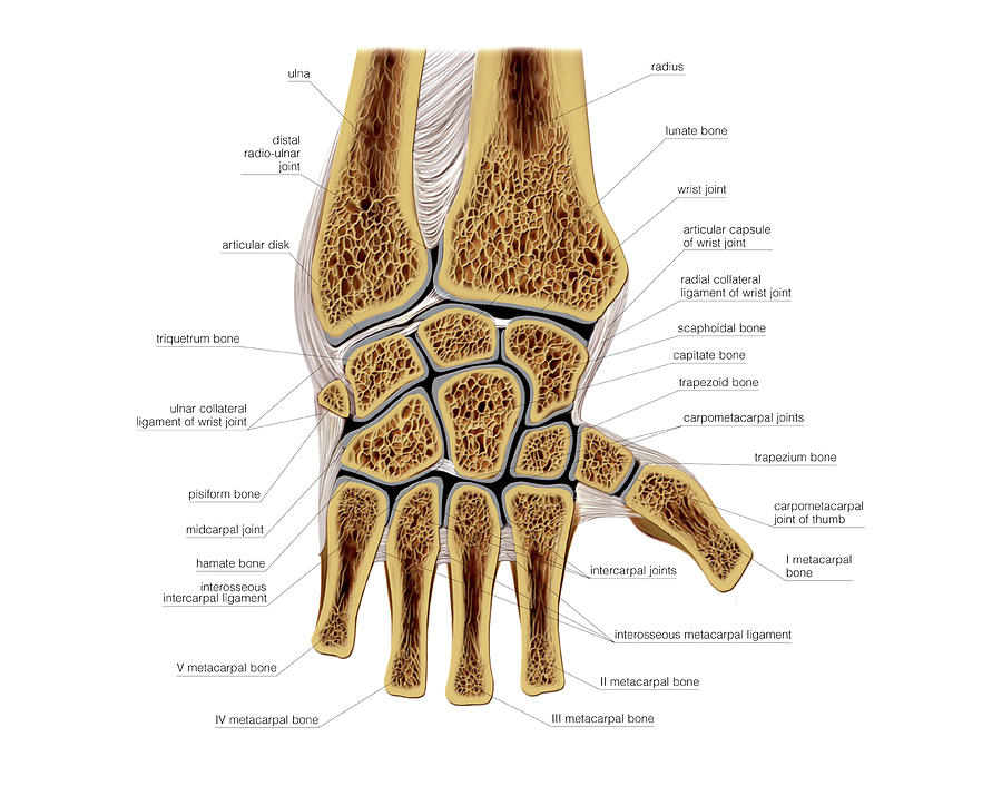
Introduction. The wrist includes three joints: the distal radioulnar joint, the radiocarpal joint and the midcarpal joint. The movements at the wrist are flexion and extension, radial and ulnar deviation and pronation and supination (at the distal radioulnar joint). Optimal wrist function requires adequate range of motion.
Tendon Diagram Of Hand / Wrist Tendonitis An Overview / Fundamentals of hand therapy, 2007.
For a minor wrist injury, apply ice and wrap your wrist with an elastic bandage. Preparing for your appointment. Although you may initially consult your family health care provider, you may receive a referral to an orthopedic surgeon, a doctor who specializes in joint disorders, called a rheumatologist, or a doctor specializing in sports medicine.

Joints of the wrist and hand Osmosis
Wrist dislocations can occur at the radiocarpal joint, the midcarpal joint, the distal radioulnar joint or may represent a combination of these injuries in severe trauma. Carpal instability in the form of lunate and perilunate dislocations are uncommon injuries but can be frequently missed. Distal radioulnar joint dislocations are most commonly associated with distal third radius fractures.

Anatomy Of The Human Wrist
Official sqadia.com Website: 🌐 https://www.sqadia.com/catalog🎬 5500+ Medical Videos☛📄 DESCRIPTIONDid you know that the wrist joint is one of the major joi.
Wrist Joint Anatomy, Bones, Carpal Tunnel Geeky Medics
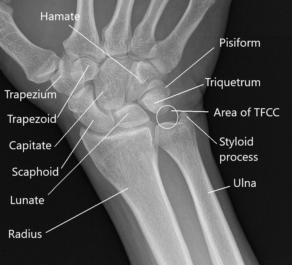
The radiocarpal joint is a synovial joint formed between the radius, its articular disc and three proximal carpal bones; the scaphoid, lunate and triquetral bones. Technically, the radiocarpal joint is considered to be the only articular component of the wrist joint; many references, however, may also include adjacent joints, such as the carpal.
Causes and Management of Wrist Joint Pain Complete Orthopedics
TRANSCRIPT. Now let's look at the wrist joint. Though we often speak of it as one joint, there are really two joints here, very close together. They're called the radiocarpal joint, and the mid-carpal joint. To understand them let's look at the bones. We'll look at them this way up. Eight small carpal bones form the carpus.
WRIST JOINT Samarpan Physiotherapy Clinic
Anatomi Wrist Joint atau Sendi Pergelangan Tangan Manusia. Sendi pergelangan tangan atau juga dikenal sebagai sendi radiokarpal adalah sendi sinovial di ekstremitas atas, menandai area transisi antara lengan bawah dan tangan. Maka dari itu artikel ini telah menuliskan bahasan dari anatomi wrist joint atau sendi pergelangan tangan manusia.

Joints of the wrist and hand Osmosis
distal radioulnar joint (Galeazzi fracture-dislocation) lunate/perilunate dislocation. associated ligamentous injury. scapholunate dissociation. Radiographic features. Diagnosis usually only requires a standard wrist x-ray series. In some complex cases, additional cross-sectional imaging (usually CT) is required to accurately assess the fracture.
Wrist Joint Anatomy Concise Medical Knowledge
Carpal bones in the wrist. Your wrist is made up of eight small bones called the carpal bones, or the carpus. These irregularly shaped bones join your hand to the two long forearm bones: the.
Wrist Joint Anatomy
Bookshelf ID: NBK534779 PMID: 30521200. The wrist joint also referred to as the radiocarpal joint is a condyloid synovial joint of the distal upper limb that connects and serves as a transition point between the forearm and hand. A condyloid joint is a modified ball and socket joint that allows for flexion, extension, abduction, and adduction.

Wrist joint (Radiocarpal joint) Medically
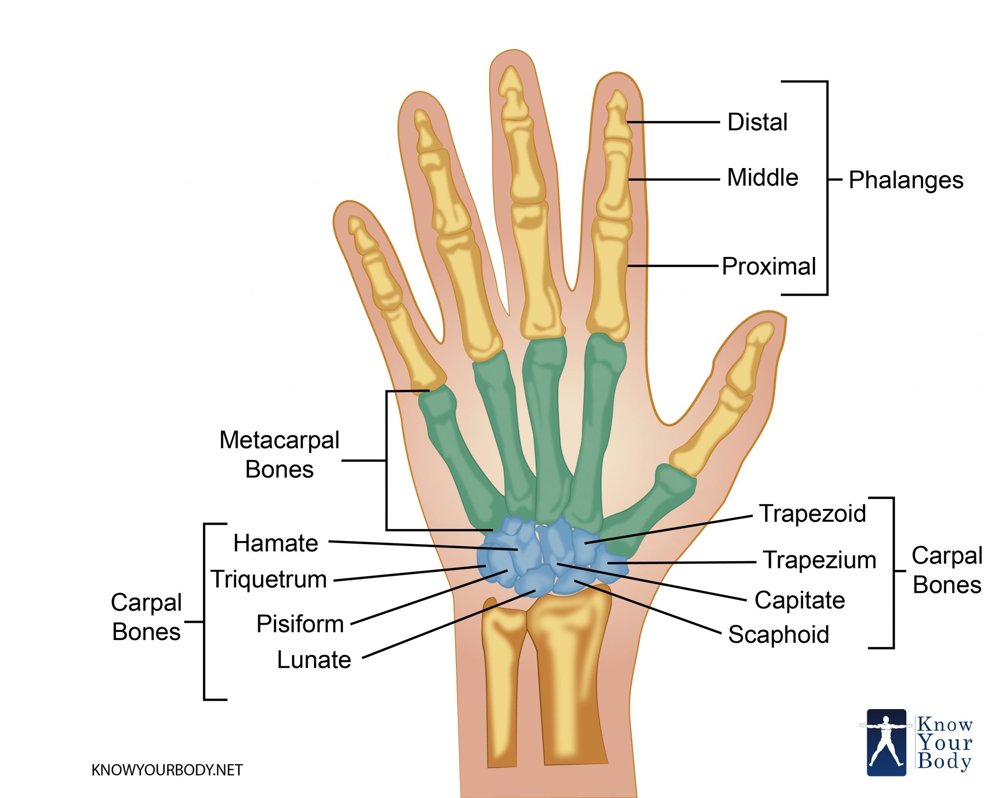
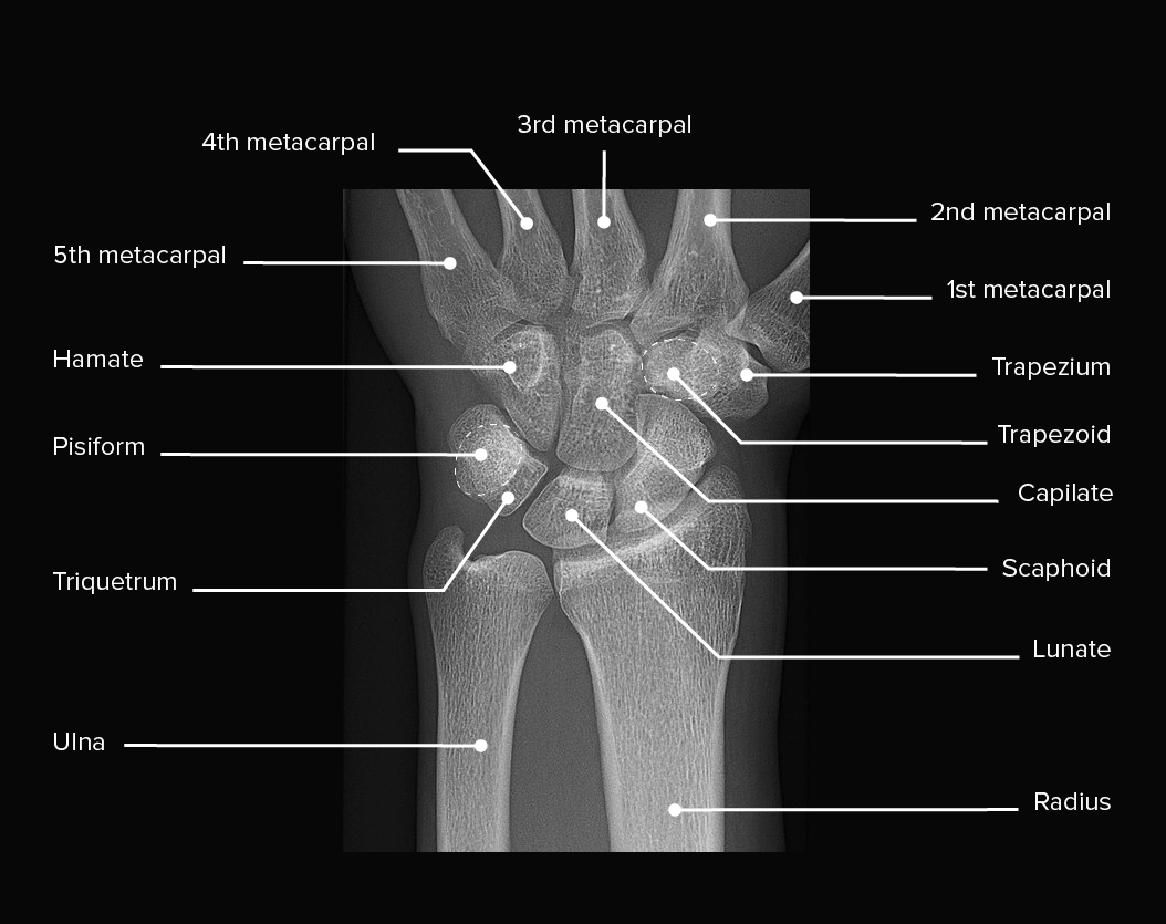
Wrist bones. Your wrist is a complex joint made of eight bones that are arranged into two rows. The proximal row (on the back of your hand, closest to your forearm) includes the: Scaphoid. Lunate. Triquetrum. Pisiform. The distal row (on the underside of your wrist closest to your palm) includes the: Trapezium.
Wrist Joints Photograph by Asklepios Medical Atlas Pixels
2.1. Bones. The wrist joint is a diarthrodial joint and is built up of eight unique carpal bones. They are interposed between the forearm (radius and ulna) and the five metacarpal bones (Figure 1).The wrist is composed of two rows of carpal bones: the proximal carpal row (PCR) includes from radial to ulnar the scaphoid, lunate, triquetrum, and pisiform; the distal carpal row (DCR) includes.

Wrist Joint AnatomyBones, Movements, Ligaments, Tendons Abduction, Flexion
The hand and wrist have a total of 27 bones arranged to roll, spin and slide [5]; allowing the hand to explore and control the environment and objects. The carpus is formed from eight small bones collectively referred to as the carpal bones. The carpal bones are bound in two groups of four bones: the pisiform, triquetrum, lunate and scaphoid on.

Wrist X Ray Anatomy The Anatomy Stories
Arthritis in Wrist. People with wrist arthritis have wrist pain, swelling and stiffness. Rheumatoid arthritis, osteoarthritis and wrist injuries can cause inflammation in the wrist joint. Steroid shots, wrist splints and anti-inflammatory drugs can ease pain and swelling. Rarely, people need surgery to improve their range of motion and reduce pain.
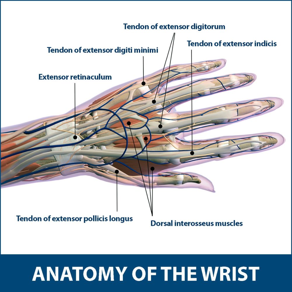
Wrist Tendonitis Florida Orthopaedic Institute
Many wrist injuries (such as fractures, also known as a broken bone) involve the joint surface. There are three joints in the wrist: Radiocarpal joint: This joint is where the radius, one of the forearm bones, joins with the first row of wrist bones (scaphoid, lunate, and triquetrum). Ulnocarpal joint: This joint is where the ulna, one of the.