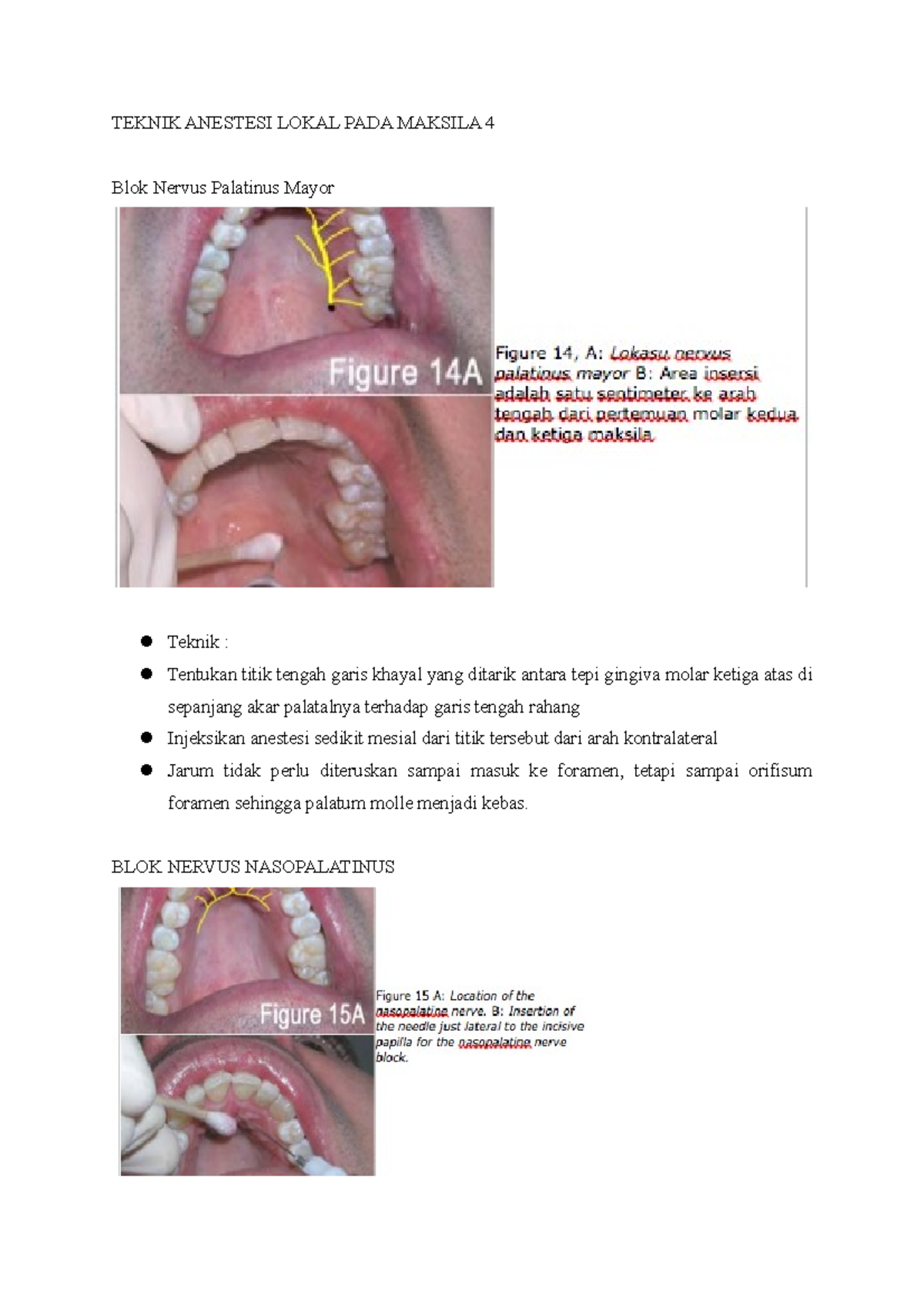
Teknik Anestesi Lokal Pada Maksila 4 TEKNIK ANESTESI LOKAL PADA MAKSILA 4 Blok Nervus
Extraction of teeth is the most common minor surgical procedure performed. Complication of extraction ranges from periodontal injury to fracture of jaw in the mandible and fracture of tuberosity and oroantral communication in the maxilla. Subconjunctival.
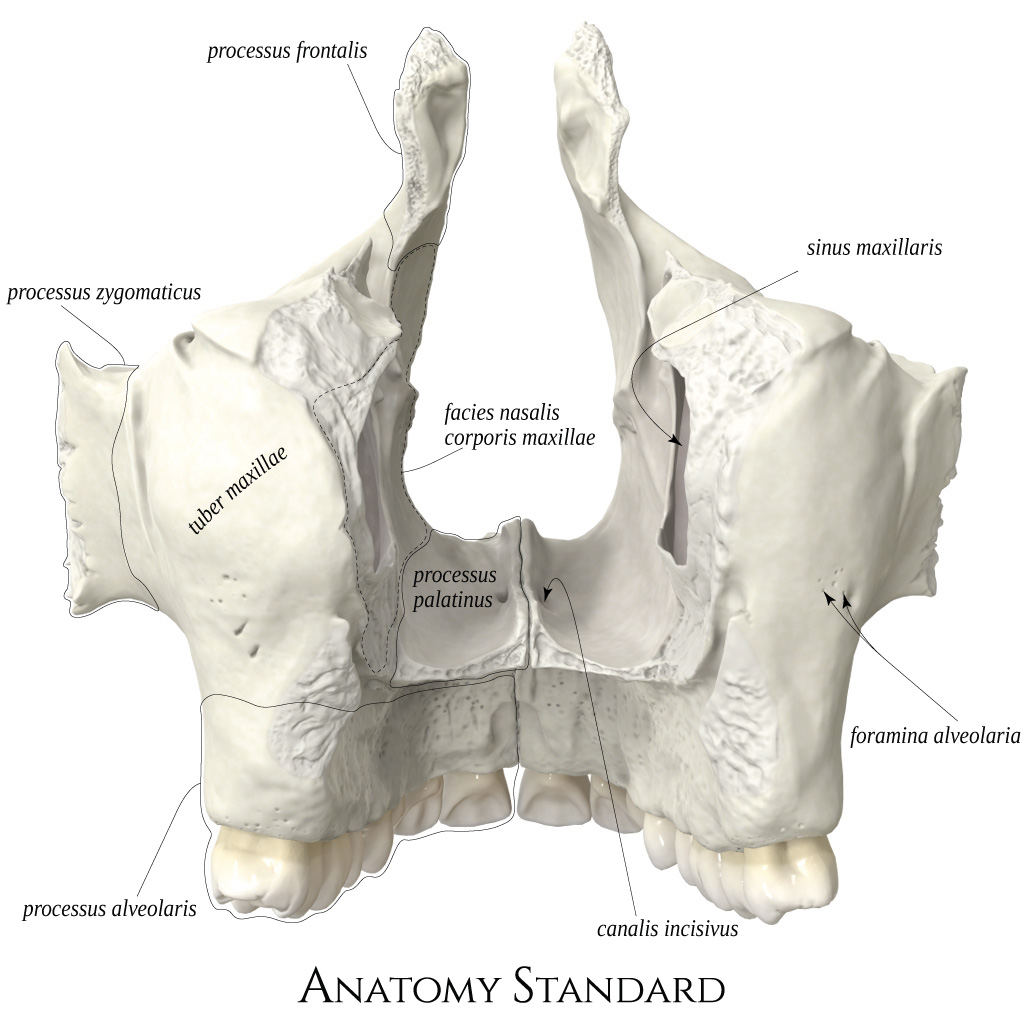
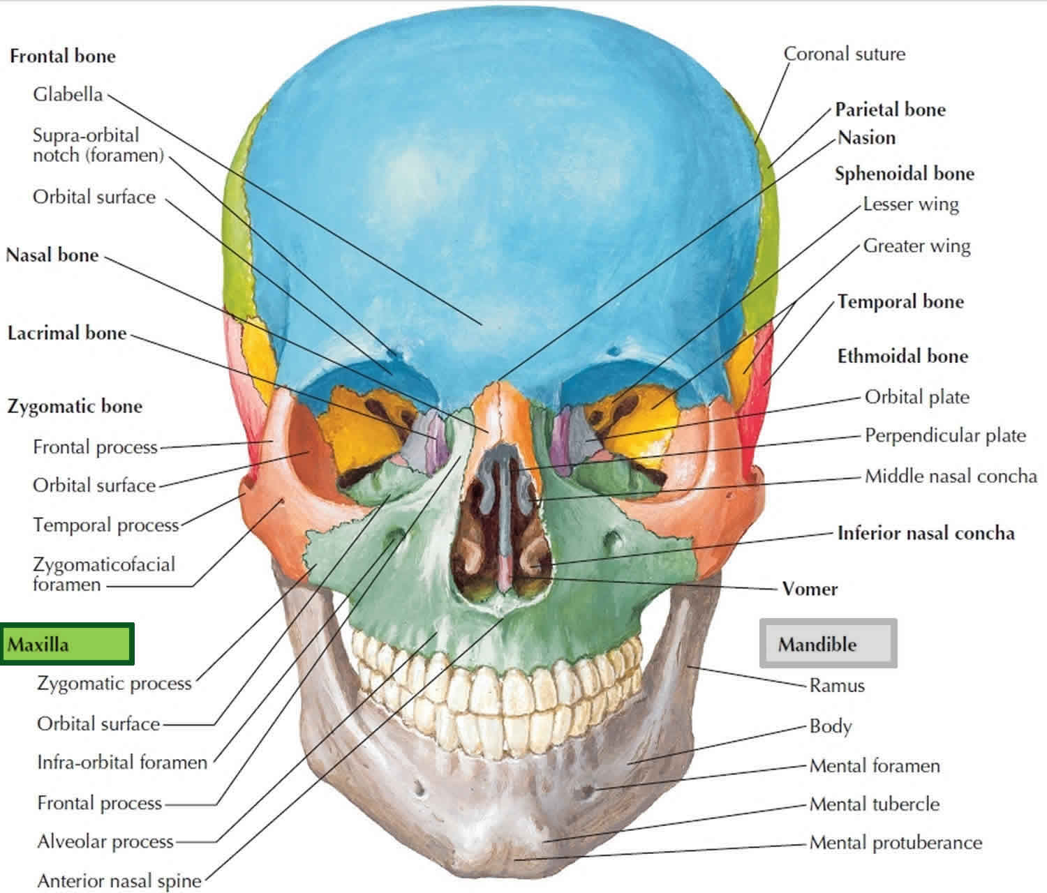
Maxilla
Maxillary fractures are one of the most common emergencies presenting in the acute setting [1]. Due to the complex anatomy within this region and the proximity to vital structures, including the brain, early diagnosis and precise treatment planning are of paramount importance. Numerous treatment methods are well-practiced globally, and these aim to restore the patient's quality of life.

2179 92102 MacamMacam Kelainan Maksila, Mandibula, Dan TMJ PDF
tuber maxilla yang lebih besar daripada ukuran normal yang tentu saja akan menyulitkan pada tahap penyusunan gigi dan juga berpengaruh pada retensi, stabilitas, dan kenyamanan. Tuber maksila besar, Material Literature Review Studi Literatur. 38 Endang Kusdarjanti, et al. | Journal of Vocational Health Studies 03 (2019): 37-39
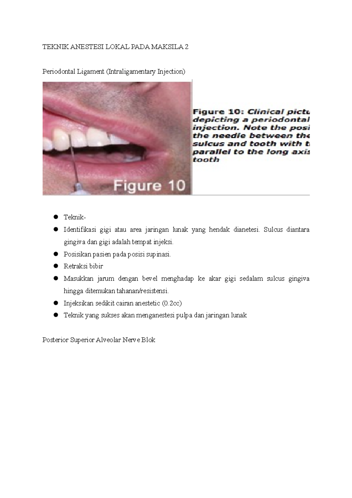
Teknik Anestesi Lokal Pada Maksila 2 TEKNIK ANESTESI LOKAL PADA MAKSILA 2 Periodontal Ligament
Tuber Maxilla adalah sebuah tonjolan yang biasanya berbentuk bulat pada posterior rahang atas akibat pertumbuhan molar 3 yang ditutupi jaringan lunak dan secara anatomi sangat penting untuk retensi, stabilitas, fungsional, dan kenyamanan pada pasien.Tujuan: Adalah untuk menjelaskan teknik pembuatan gigi tiruan lengkap lepas dengan tuber maxilla.

Fraktur Mandibula & Maksila PDF
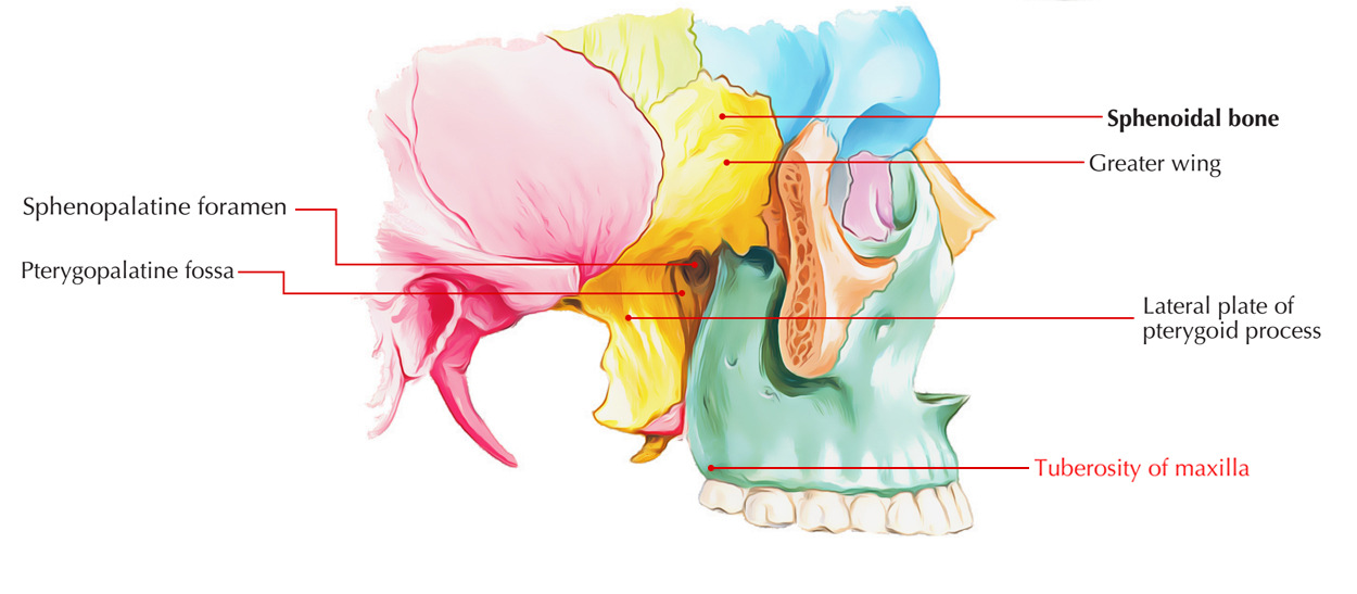
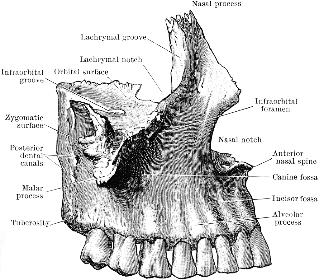
Definition. T he maxillary tuberosity is at the lower part of the infratemporal surface of maxilla. It is a rounded eminence, especially prominent after the growth of the wisdom tooth; it is rough on its lateral side for articulation with the pyramidal process of the palatine bone and in some cases articulates wth the lateral pterygoid plate of.
99 de Motive Trufele de Romania
The maxilla (pl.: maxillae / m æ k ˈ s ɪ l iː /) in vertebrates is the upper fixed (not fixed in Neopterygii) bone of the jaw formed from the fusion of two maxillary bones. In humans, the upper jaw includes the hard palate in the front of the mouth. The two maxillary bones are fused at the intermaxillary suture, forming the anterior nasal spine.This is similar to the mandible (lower jaw.

Fractured Maxillary Tuberosity Exodontia
Kasus: Rahang atas tidak bergigi dengan kasus tuber maksila yang besar pada posterior. Tinjauan pustaka: Single complete denture merupakan gigi tiruan lengkap dimana antagonisnya dapat berupa gigi asli baik sebagian maupun keseluruhannya. Sedangkan complete denture merupakan gigi tiruan lengkap rahang atas dan rahang bawah yang digunakan jika.
Maxillary Tuberosity (Tuberosity of Maxilla) Earth's Lab
Anatomical terms of bone. [ edit on Wikidata] At the lower part of the infratemporal surface of the maxilla is a rounded eminence, the maxillary tuberosity, especially prominent after the growth of the wisdom tooth; it is rough on its lateral side for articulation with the pyramidal process of the palatine bone and in some cases articulates.
Outer View of Superior Maxilla ClipArt ETC
Tuber maksila Kanan : besar/kecil Kiri : besar/kecil Daerah ini ditutup oleh jaringan fibrosa dengan ketebalan yang berbeda- beda. Disebut kecil bila ukuran tuber lebih kecil dari prosesus alveolar dan besar bila tuber melebar atau menonjol ke arah oklusal atau lateral. Tuber yang besar dapat mengganggu retensi gigi tiruan.
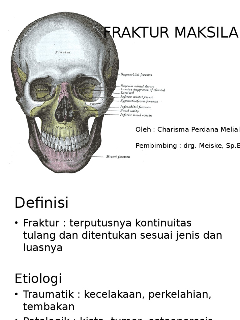
FRAKTUR MAKSILA
Hello lovely people, Link to posterior palatal seal video 👇https://youtu.be/6IyKO0mkibk

Maxilla. Lateral view Anatomy bones, Human anatomy and physiology, Skull anatomy
The tuber maxillae medially articulate with the pyramidal process of the palatine bone and, in the atrophic maxilla, often reduce this bone amount and height because of sinus pneumatization. Posteriorly, the maxillary tuber joins the pterygoid process of the sphenoid bone forming the pterygomaxillary junction. Cranially, at the end of the.

Tuber melanosporum, Vittadini 1831 (Fungi Peziz… Flickr
The maxilla is a bone which helps to make up the skull. It is specifically located in the mid face, forms the upper jaw, separates the nasal and oral cavities, and contains the maxillary sinuses (located on each side of the nose. One of the maxilla's most important functions is to make up the architecture of our faces and to support the rest of.
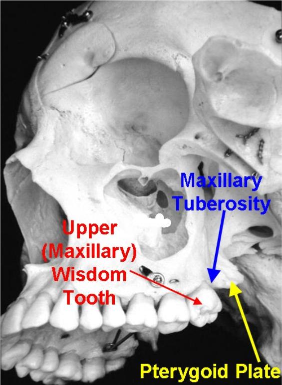
Maxillary Tuberosity Fracture
Scribd is the world's largest social reading and publishing site.

Sobo 1909 100 Palatine process of maxilla Maxilla Wikipedia Dental anatomy, Palatine
maksila besar, Material. Literature Review.. Conclusion: Tuber maxillae and Spina nasalis anterior have stable dimensions. The areas of canine and premolars are varied, because the zone is.
Maxilla bone, maxilla anatomy & maxilla function
Articular tubercle. The articular tubercle ( eminentia articularis) is a bony eminence on the temporal bone in the skull. It is a rounded eminence of the anterior root of the posterior end of the outer surface of the squama temporalis. This tubercle forms the front boundary of the mandibular fossa, and in the fresh state is covered with cartilage.

Tuber maxillae Anatomy for artists, Illustration, Lion sculpture
Introduction. The maxilla is the most important bone of the midface. It has a central location and provides structural support to the viscerocranium. It has functional and aesthetic significance as it has a fundamental role in facial architecture, separates the nasal and oral cavities, forms the upper jaw, and contains the maxillary sinus. [1] [2]