Chest XRay Projection Chest XRay MedSchool
Dari hasil pemwriksaan thorax foto PA view, errect, simetris, inspirasi dan kondisi cukup.

Perbedaan Foto Thorax Ap Dan Pa
Nov 28, 2017 Membaca foto thoraks X-Ray adalah kompetensi penting dokter umum. Banyak kelainan penyakit yang dapat dikonfirmasi dengan foto thoraks PA saja. Misalnya pada pasien dengan tuberkolosis paru, gagal jantung kronik atau pneumonia. Sehingga penting bagi dokter umum untuk dapat menguasai teknik dasar membaca foto thoraks X-Ray.
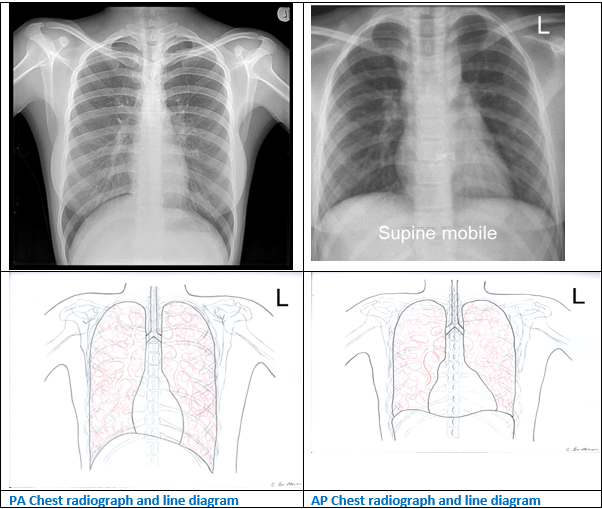
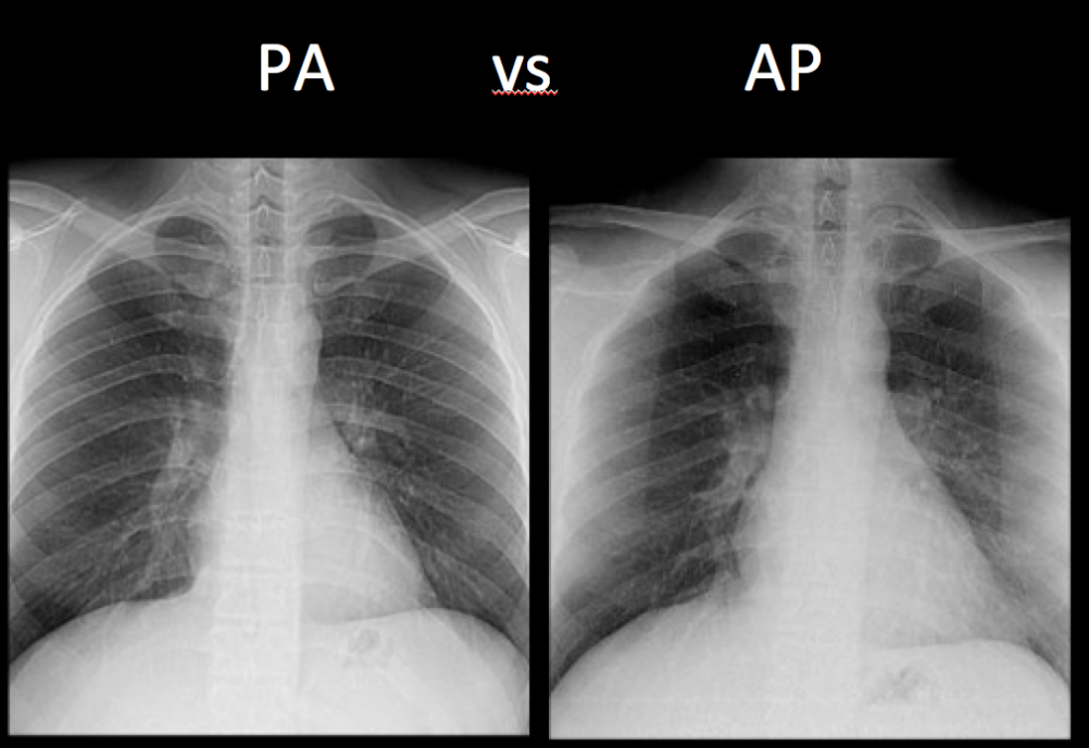
The difference between Chest Posterior Anterior (PA) and Anterior Posterior (AP) radiographs.
Study design. A cross-sectional, 3-phase study was conducted retrospectively. In phase 1, by manipulating chest CT to simulate chest PA and AP at different radiation distances, we determined CD Chest PA /CD Chest AP ratios in terms of the radiation distance. If the ratio is fixed at a specific distance to perform chest AP, we would be able to infer CD Chest PA by multiplying CD Chest AP by.

Difference between Chest AP & PA Chest PA Vs AP By BL Kumawat YouTube
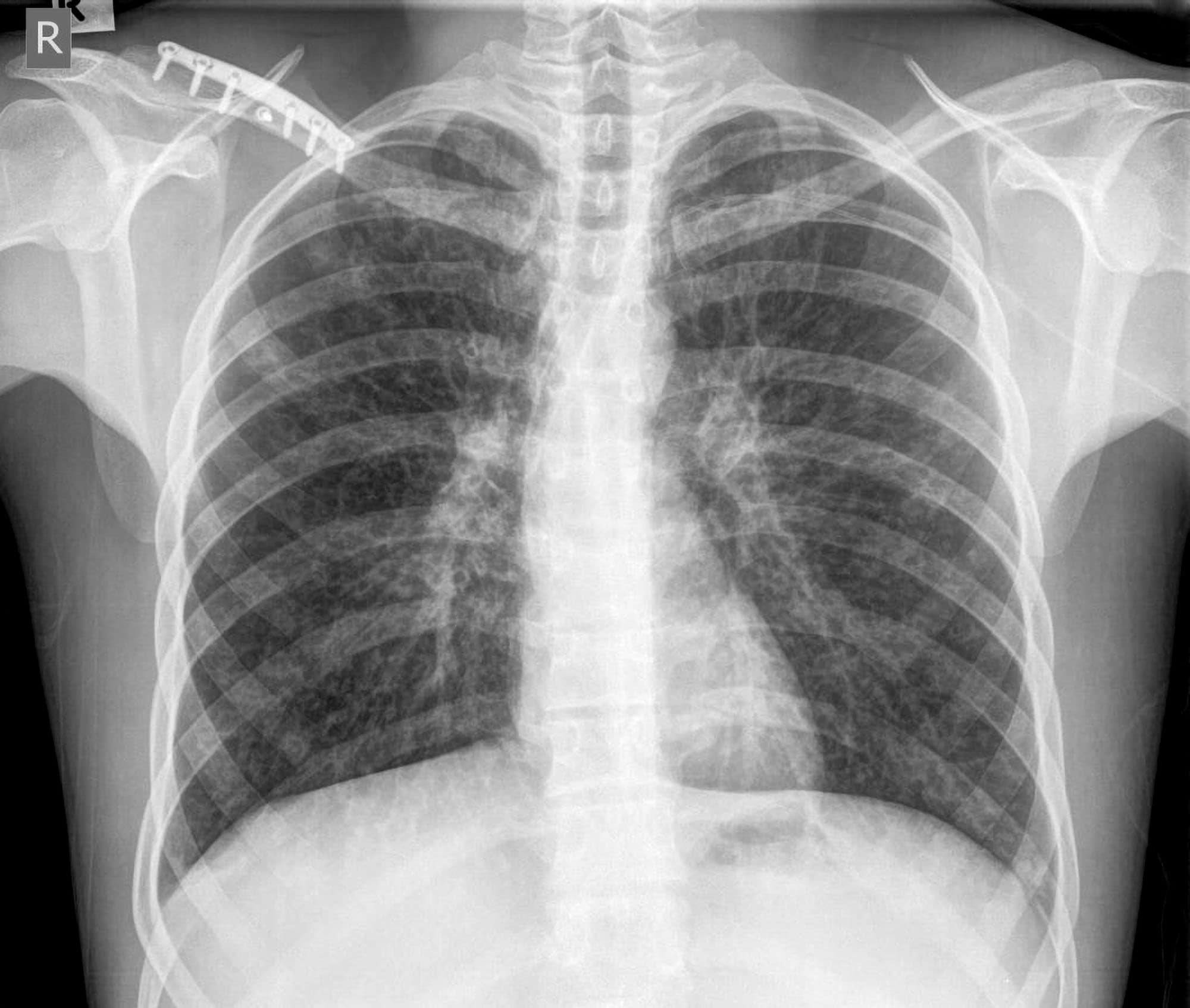
The answer is… below. On the PA view, the cardiac borders are smaller and more defined. Given the way the x-ray beam works, the heart appears smaller and with sharper borders on the PA view. The reason is that the patient's chest (anterior) is against the x-ray film with the beam entering from posterior (P) to anterior (A) - hence the.
Teknik Rontgen Toraks Alomedika
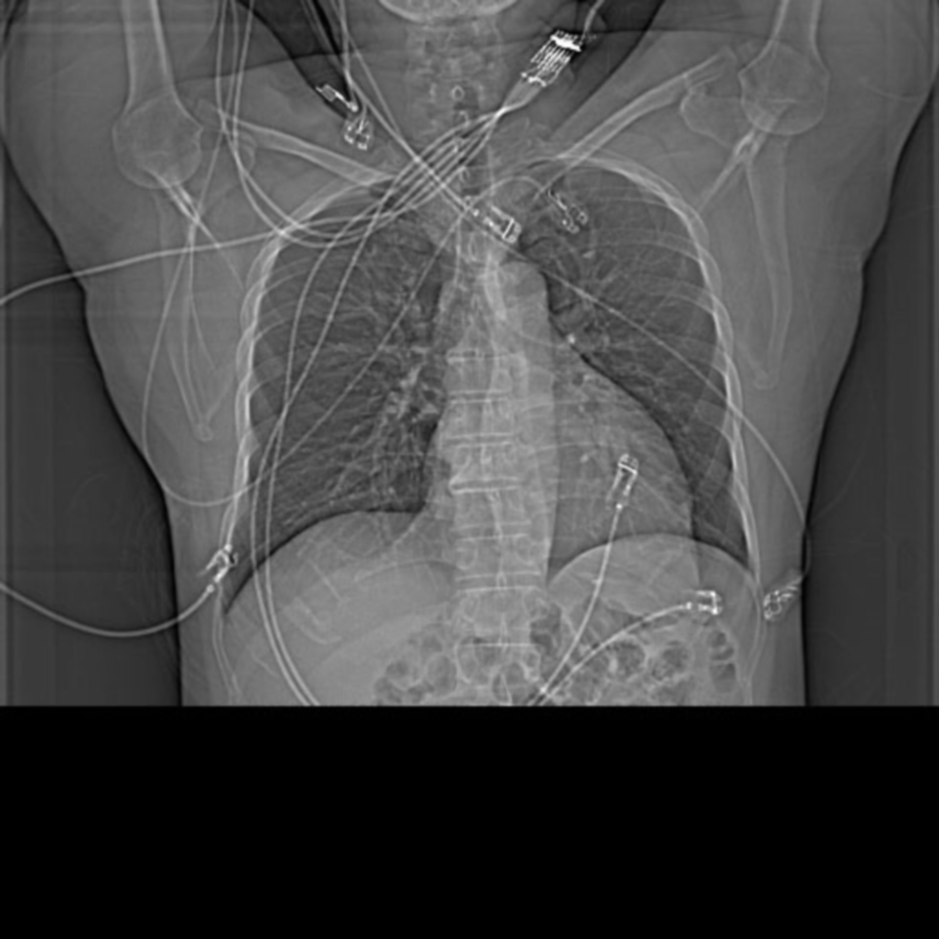
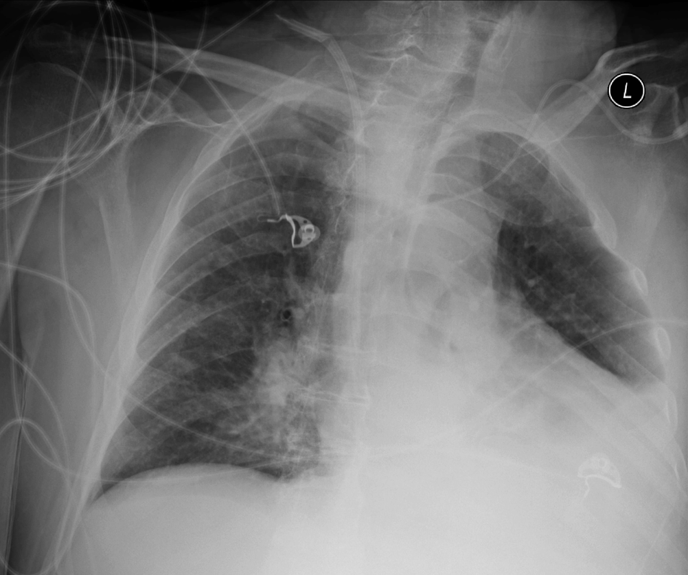
Indications. The erect anteroposterior chest view is an alternative to the PA view when the patient is too unwell to tolerate standing or leaving the bed 1.The AP view examines the lungs, bony thoracic cavity, mediastinum, and great vessels.This particular projection is often used frequently to aid diagnosis of acute and chronic conditions in intensive care units and wards.

PA VIEW Vs AP VIEW CHEST X RAY YouTube
Foto: rontgen dada (healthline.com) Saat pandemi COVID-19 terjadi, pemeriksaan thorax menjadi salah satu prosedur yang sering dianjurkan dokter untuk memeriksa kondisi paru-paru pasien yang terinfeksi virus corona.. Hal ini karena COVID-19 menyerang saluran pernapasan seseorang. Namun, ternyata tidak hanya kondisi COVID-19 saja yang membutuhkan pemeriksaan thorax atau rontgen dada.
Röntgen Thorax, a.p. DocCheck
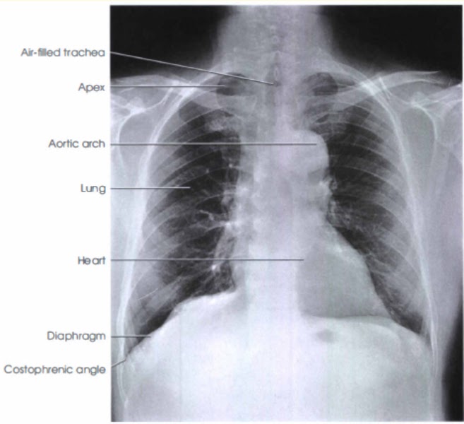
Rontgen dada atau rontgen thorax adalah foto dada yang menunjukkan jantung, paru-paru, saluran pernapasan, pembuluh darah, dan nodus limfa Anda. Rontgen dada juga bisa menunjukkan tulang belakang dan dada, termasuk tulang rusuk, tulang selangka, dan bagian atas tulang belakang Anda.
Chest PA View Radiology Basics Radtechonduty
pemeriksaan THORAX AP / PA -Tampak infiltrat pada daerah parahiler -Hilus lebar -Cor normal -Sinus dan diafragma baik. kesan : -TB paru. mohon penjelasannya ya dok,dan tindakan apa yang harus saya lakukan. terimakasih Dijawab oleh: dr. Aldo Ferly Dokter 23 Februari 2016 11:48

dokter chandra
Rontgen Dada/Thorax AP/PA Perempuan & laki-laki Semua Pemeriksaan radiologi bagian dada untuk menilai kondisi paru-paru, tulang rusuk, ukuran jantung, serta organ lainnya di dalam rongga dada Pemeriksaan ini dapat menunjang diagnosis: - Kelainan jantung bawaan, gagal jantung, dan masalah jantung lainnya - Radang paru-paru - Emfisema

PA Chest Projection. Radiology student, Medical knowledge, Radiography
1. Radang Paru-Paru Radang paru-paru, atau yang dikenal dengan istilah pneumoniaadalah infeksi bakteri atau virus yang menyerang kantung udara (alveoli). Infeksi tersebut memicu penumpukan cairan di dalam paru-paru, sehingga kapasitas organ dalam menampung oksigen menjadi berkurang.
Chest XRay Basics PA vs. AP radRounds Radiology Network
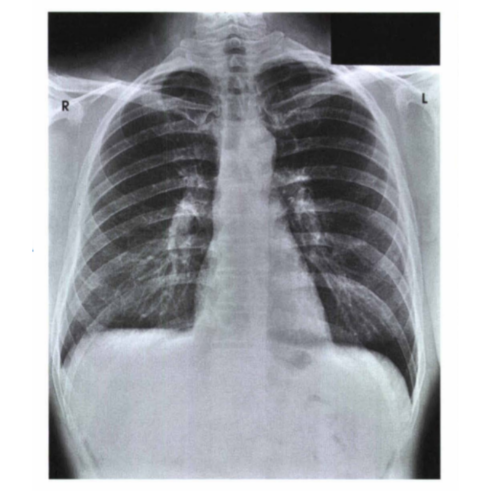
5. The chest radiograph assessement. 1. The difference between Chest Posterior Anterior (PA) and Anterior Posterior (AP) radiographs. Erect PA projections are considered the 'gold standard' for chest projection imaging (CXR). In some instances, it will not be possible to acquire an erect PA or even an erect AP image and the radiographer.
Gambaran radiograf terhadap posisi anatomi
Foto thorax AP (Anteroposterior) dan PA (Posteroanterior) adalah dua jenis teknik yang umum digunakan untuk mendapatkan gambaran struktur dada dan organ-organ yang terdapat di dalamnya, terutama paru-paru.

PA vs AP view... Medical radiography, Radiology technician, Medical ultrasound
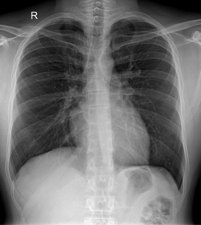
The chest x-ray is the most common radiological investigation in the emergency department 1. The PA view is frequently used to aid in diagnosing a range of acute and chronic conditions involving all organs of the thoracic cavity. Additionally, it serves as the most sensitive plain radiograph for the detection of free intraperitoneal gas or.

Is this an AP or PA chest xray? r/Radiology
Thorax ap adalah singkatan dari anterior-posterior, yang berarti foto diambil dari belakang ke depan. Di foto ini, posisi kamu mungkin tampak seperti sedang membuat pose pahlawan super! Tapi jangan khawatir, kamu tidak perlu mengenakan jubah super untuk mendapatkan foto ini.

PA vs AP view Chest radiology YouTube
Age: 30. Gender: Male. x-ray. The superior mediastinum appears widened due to AP magnification. The heart appears enlarged - a combination of AP magnification and underinflation. There appears to be a bilateral interstitial infiltrate/airways thickening - probably also due to underinflation. x-ray. Same patient, repeat CXR the next day (PA.
The difference between Chest Posterior Anterior (PA) and Anterior Posterior (AP) radiographs
Peralatan Beberapa peralatan yang umumnya digunakan dalam pemeriksaan thorax, antara lain x-ray generator, bucky stand x-ray dengan bucky protector dan faceplate, serta mesin portable x-ray yang dapat dibawa ke tempat tidur pasien di Rumah Sakit dan ruang gawat darurat. [1,10] X-ray Generator