Causes and Management of Wrist Joint Pain Complete Orthopedics
Berdasarkan pengamatan penulis dalam mengamati seluruh teknik pemeriksaan Wrist Joint di dalam jurnal George D. Xipoleas, dkk (2014), jurnal N. Sifi, dkk(2019) dan Anil K. Bhat, dkk (2011) sudah baik mempunyai alasan tersendiri dalam pengambilan foto radiografi sebaiknya proyeksi yang digunakan dalam pemeriksaan. Twitter;

Joints of the wrist and hand Osmosis
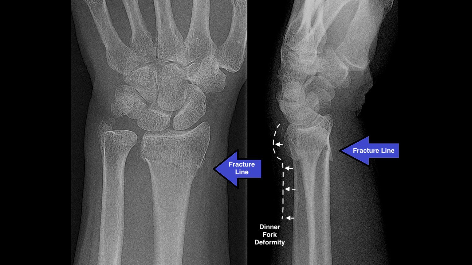
Hasil penelitian menunjukkan bahwa teknik pemeriksaan wrist joint proyeksi ulnar deviation pada kasus fraktur scaphoid yaitu menyiapkan kaset ukuran 18 x 24 cm dengan pasien duduk di meja pemeriksaan, posisi objek tangan berada diatas IR, posisi tangan PA dengan wrist joint dirotasikan atau difleksikan
Wrist Xray Interpretation OSCE Guide Geeky Medics
The CT hand and wrist protocol serves as an examination for the bony assessment of the wrist and is often performed as a non-contrast study and less often as a contrast-enhanced study. A CT wrist can be also conducted as a CT arthrogram for the evaluation of ligamentous injuries and the triangular fibrocartilage complex.. Note: This article aims to frame a general concept of a CT protocol for.
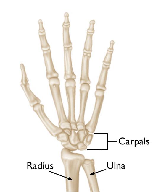
bones of wrist joint
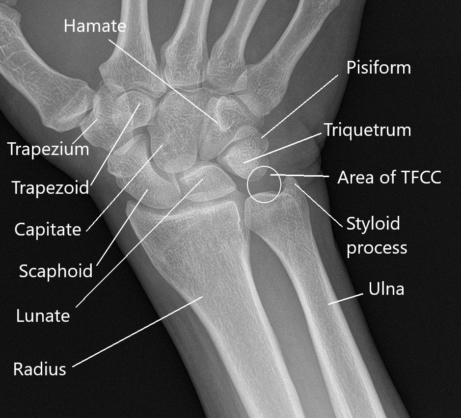
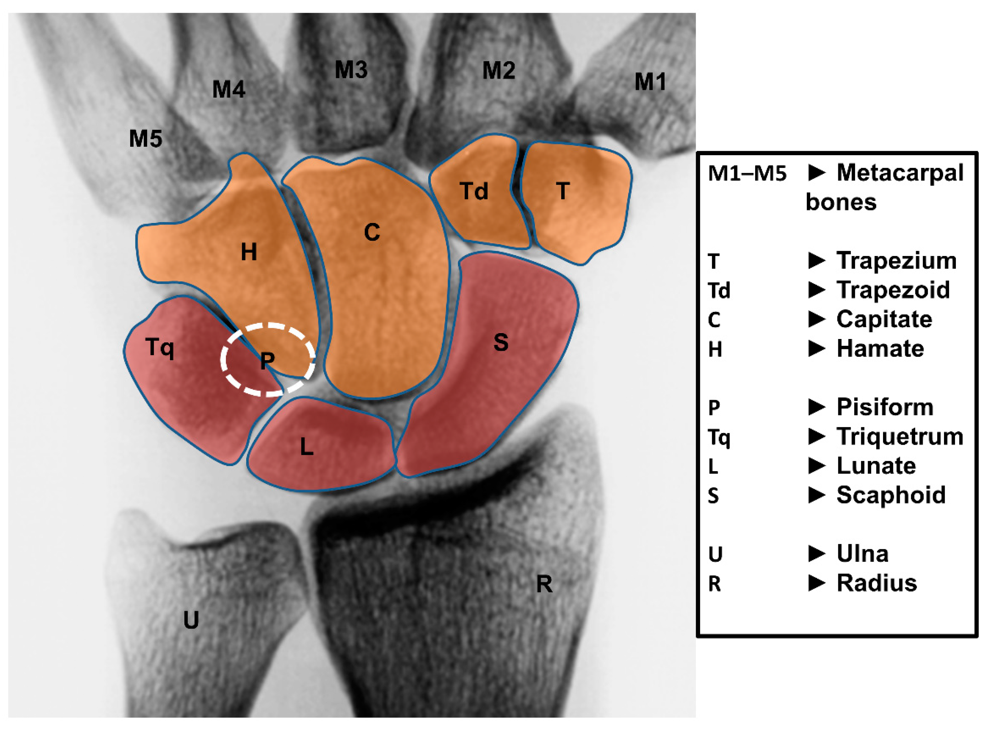
Proyeksi Pemeriksaan Wrist Joint. Tetapi untuk Proyeksi pemeriksaan di lapangan yaitu PA dan LATERAL untuk Proyeksi-proyeksi diatas bisa dilihat dari basic radiologi yang ada di buku. PP = Pasien duduk di meja pemeriksaan dengan antebrachi di fleksikan dan telapak tangan menempel pada kaset. Kriteria gambaran = Metacarpal, Carpal ( Schapoid.

Proyeksi Pemeriksaan Wrist Joint PDF
40 JURNAL RADIOGRAFER INDONESIA, ISSN 2620-9950 PROSEDUR PEMERIKSAAN MRI WRIST JOINT PADA KASUS DISRUPSI DISTAL RADIOULNAR JOINT DENGAN MENGGUNAKAN GENU COIL Hendriawan1), M.Irwan Katili2), Dartini3) 1, 2,3) Poltekkes Kemenkes Semarang Author Mail : [email protected]
Wrist Joint Replacement (Wrist Arthroplasty) OrthoInfo AAOS
NS Muhammad Izzudin, Hermina Sukmaningtyas. Jurnal Radiografer Indonesia 4 (2), 99-105. , 2021. 4 *. 2021. PROSEDUR PEMERIKSAAN MRI WRIST JOINT PADA KASUS DISRUPSI DISTAL RADIOULNAR JOINT DENGAN MENGGUNAKAN GENU COIL. D Hendriawan, M. Irwan Katili. Jurnal Radiografer Indonesia 2 (1), 40-47. , 2019.

Wrist and Hand Injuries Musculoskeletal Key
TEKNIK RADIOGRAFI WRIST JOINT By yumanet Wednesday, April 18, 2018 Share Tweet Share Share Email. Radiografer. Persiapan Pasien. Teknik Pemeriksaan. Proyeksi PA. Posisi Pasien : Duduk menyamping dari meja pemeriksaan. Posisi Objek : - lengan bawah menempel meja pemeriksaan - atur wrist pada pada pertengahan pasien.
Life Free FullText Anatomy, Biomechanics, and Loads of the Wrist Joint
About Press Copyright Contact us Creators Advertise Developers Terms Privacy Policy & Safety How YouTube works Test new features NFL Sunday Ticket Press Copyright.

Wrist Joint AnatomyBones, Movements, Ligaments, Tendons Abduction, Flexion
Radiologi
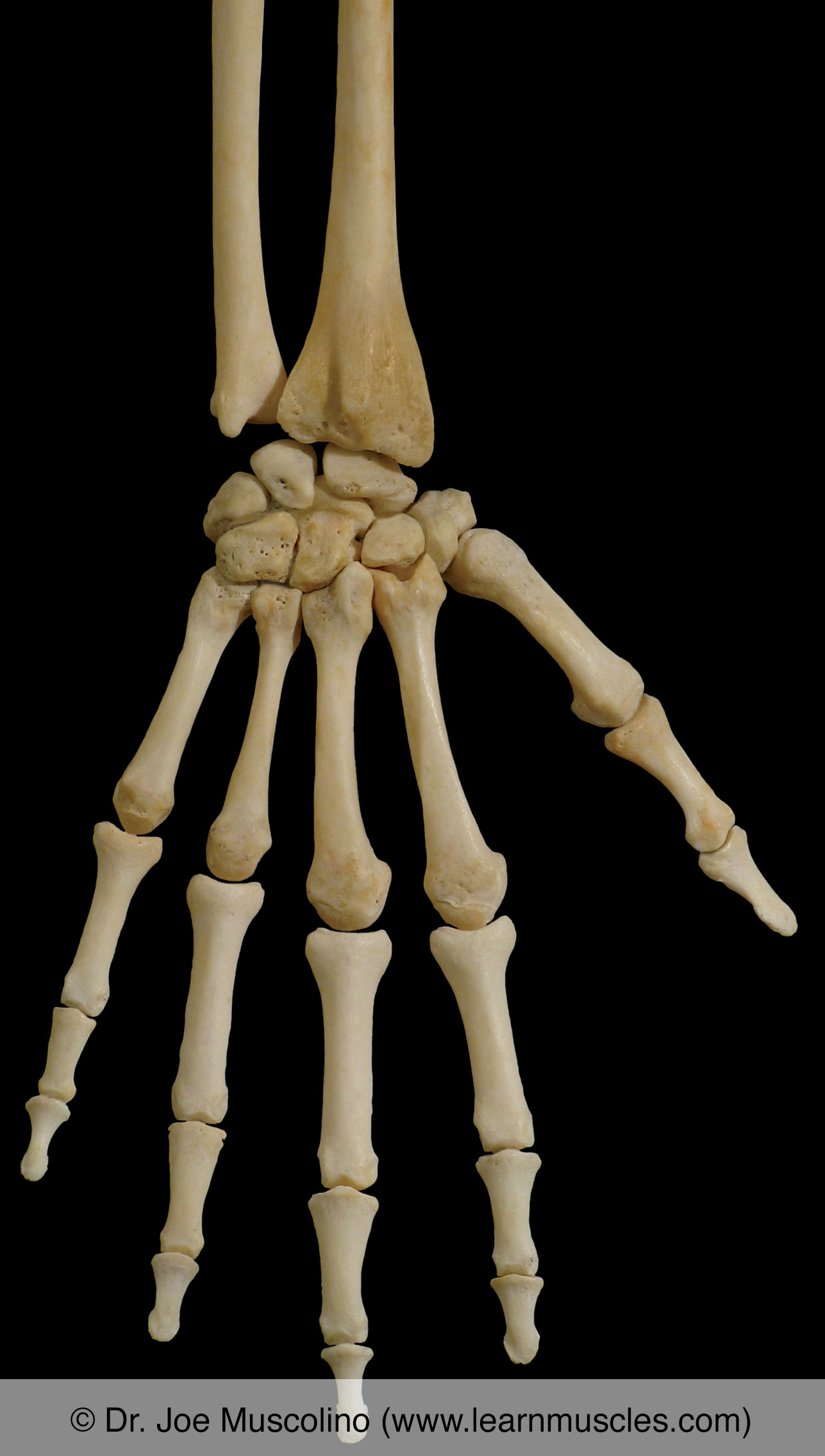
Wrist Joint Learn Muscles
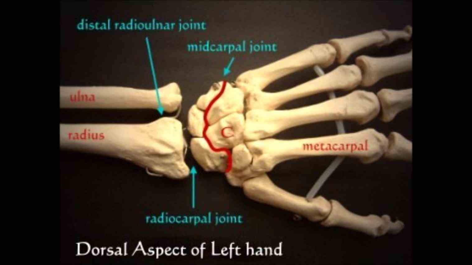
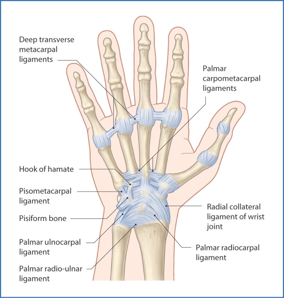
The wrist joint is a complex joint consisting of several bones and joints.. memposisiskan pasien dengan cara lengan disamping tubuh, tangan dan wr ist. Coronal pada pemeriksaan MRI Wrist Joint .
Arthritis of the Wrist OrthoInfo AAOS
TEKNIK PEMERIKSAAN WRIST JOINT | PDF. Scribd adalah situs bacaan dan penerbitan sosial terbesar di dunia.

Simulasi komunikasi pelayanan pemeriksaan elbow joint di ruang Radiologi YouTube
ABSTRACT Background: Procedure of MRI wrist joint examination on disruption case of Distal Radioulnar Joint at radiology installation of RS Panti Rapih Yogyakarta using genu coil. The purpose of this research is to know the procedure of MRI wrist joint examination in DRUJ disruption case using genu coil, to know the reason for the use of genu coil for the examination of wrist joint and to know.
WRIST JOINT Samarpan Physiotherapy Clinic
Latar belakang: Teknik pemeriksaan wrist joint untuk melihat kelainan pada daerah carpalia khususnya pada os scaphoid ada teknik khusus yaitu ulnar deviation dengan variasi central ray 150 sampai 25 0 proximally. Tujuan: untuk mengetahui pada arah sinar yang mana untuk menilai anatomi scaphoid yang optimal.

Joints of the wrist and hand Osmosis
Teknik Pemeriksaan Radiografi Wrist Joint. Dipos oleh Rini pada Mei 17, 2021. Proyeksi : PA. Kaset : ukuran 18 x 24 cm. kV : 60 ± 6 mAs : 4. FFD : 100 cm. Posisi Pasien : Pasien duduk menyamping meja pemeriksaan,siku flexi 90°, posisi tangan dan lengan bawah berada di atas meja pemeriksaan.

Radiological imaging of the wrist joint Orthopaedics and Trauma
Teknik Radiografi Manus, Wrist joint, Antebrachii, Humerus INDIKASI PEMERIKSAAN RADIOGRAFI • Trauma / cidera - Fraktur, fisura, dislokasi, luksasi, ruptur • Pathologis - Artheritis, Osteoma, dll. • Benda asing (corpus alienum) • Cacat bawaan / Congenental - Polidactile. PROSEDURE / PRINSIP PROTEKSI RADIASI Prinsip Jarak Dalam setiap pemotretan dengan menggunakan sinar-X seorang.

Wrist joint (Radiocarpal joint) Medically
Ultrasound is a useful imaging modality for evaluation of the wrist, allowing high-resolution imaging of anatomy while simultaneously allowing dynamic evaluation of the joints, tendons, and ligaments.. Approach. There are multiple possible approaches to imaging the wrist with ultrasound. The exam is easily tailored to a specific painful area or set of differential diagnoses.