
Röntgenfoto van de schedel stock afbeelding. Image of onderzoek 5803779
Schedel- en hersenletsel Inleiding. Iedereen is er zich wel van bewust dat een letsel aan het hoofd (Latijn: trauma capitis) een ernstige zaak is. In het hoofd zitten immers de hersenen, die de grondslag zijn van ons hele geestelijke leven. Omdat de hersenen in mechanisch opzicht een brijig zachte massa vormen en dus heel kwetsbaar zijn voor.

A Schedel AP Xray of a Skull Free Stock Photo by Sugiyatno on
Scribd adalah situs bacaan dan penerbitan sosial terbesar di dunia.
Schädel Röntgen Bild mit Hals Wirbelsäule seitlich vom Kind StockFoto Adobe Stock
ProZ.com Headquarters 235 Harrison Street Suite 202 Syracuse, NY 13202 USA +1-315-463-7323

Rol Van De Schedel Röntgenbeeld Van De SchedelAP Mening of Voormening Van De Schedel Stock Foto
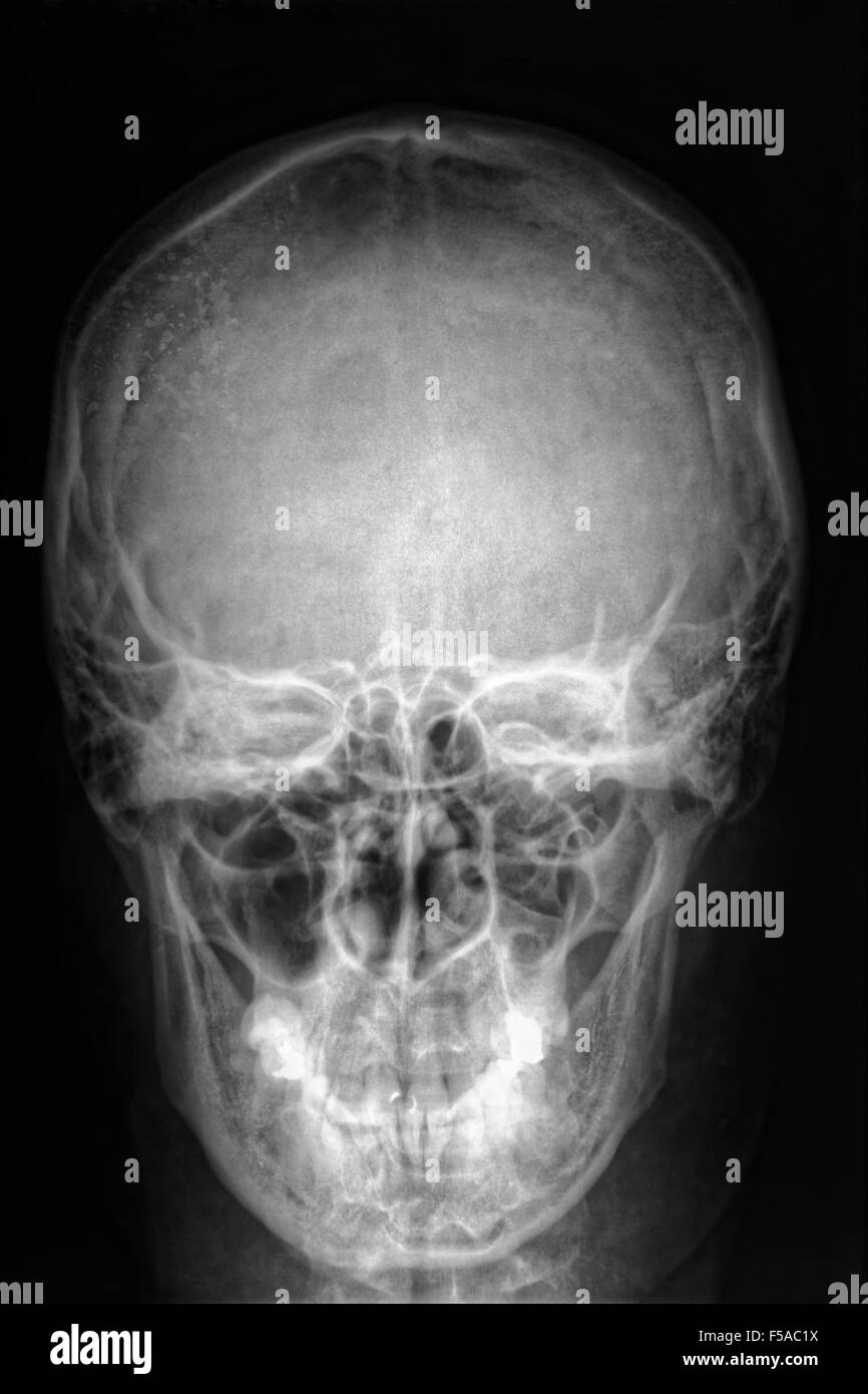
A Schedel AP radiograph of the skull. Head and Neck x ray radiology image black and white effect on black background color.. medical medicine neck neurological orthopedic patient people physical prevention radiography radiological radiologist radiology ray roentgen science skeletal skeleton skulls surgery technology therapy tomography trauma.

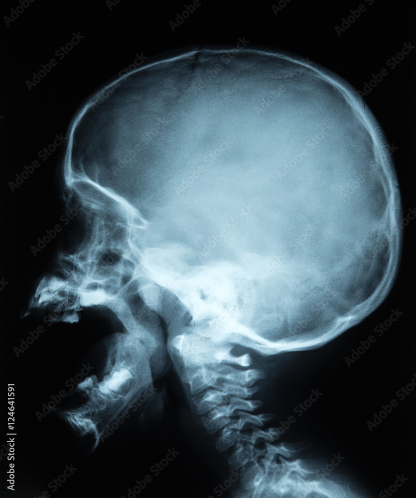
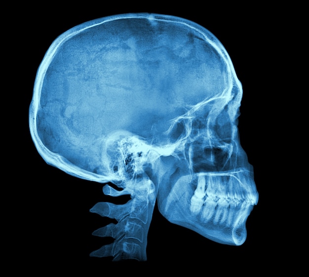
SKULL LATERAL VIEW
Balas. LUMBOSACRAL 1. anatomi lumbosacral 2. Persiapan Pasien Tidak memerlukan persiapan kusus, hanya melepas atau menyi. Teknik Pemeriksaan Schedel ( Kepala ) PROYEKSI AP POSISI PASIEN Pasien tidur pada posisi Supine di atas meja pemeriksaan, dengan MSP tubuh tepat pada Mid L. Pencucian Film Rontgen.

Röntgenfoto van de schedel stock afbeelding. Afbeelding bestaande uit röntgen 43951557
We would like to show you a description here but the site won't allow us.

Het Beeld Van De Röntgenstraal Van De Schedel Stock Foto Image of gebied, segment 18420948
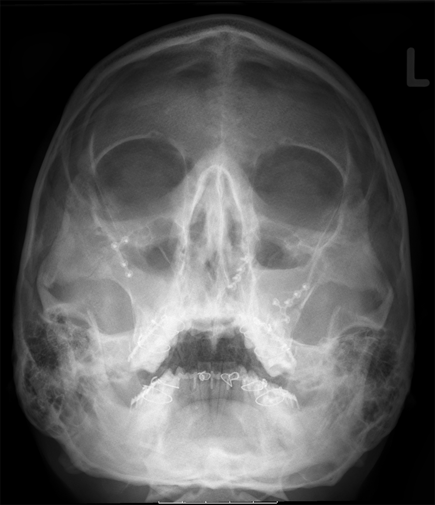
Citation, DOI, disclosures and article data. The Caldwell view is a caudally angled radiograph, with its posteroanterior projection allowing for minimal radiation to the orbits. This view may be used in imaging of the skull or facial bones depending on the clinical indications.

Schedelhoofd Medische Röntgenstraal Stock Foto Afbeelding bestaande uit hoofd, gezondheid
Pada rontgen kepala posisi Waters, idealnya piramida tulang petrosum diproyeksikan pada dasar sinus maksilaris sehingga kedua sinus maksilaris dapat dievaluasi sepenuhnya. Rontgen kepala posisi Waters biasanya dilakukan pada keadaan mulut tertutup. [5,6] Rontgen Kepala Posisi Submentoverteks.

Röntgenfoto van de schedel stock afbeelding. Afbeelding bestaande uit onderzoek 5803779
The Atlas of Normal Roentgen Variants That May Simulate Disease is a classic radiology text that was first published in 1973, and is now in its ninth edition (2012) 1,3.The first - and all subsequent - editions, were written by an American radiologist Theodore Eliot Keats (1924-2010) who died during the development of its most recent iteration 1,2..

Schedelhoofd Medische Röntgenstraal Stock Foto Image of gezondheid, ziekenhuis 74566508
Schedel rontgen lateral showed there were suspected linear fracture os frontal dextra. Ophthalmologic examination revealed uncorrected visual acuity (UCVA) was 0.25 eccentric view on RE and 1.0 on left eye (LE). Ocular motility were full and intraocular pressure (IOP) using noncontact tonometry (NCT) is 22

Röntgenstraal Van Misvormde Schedel Stock Foto Afbeelding bestaande uit gebroken, fysiek 2378114
After the nasal bones, the mandible is considered the second most common site of facial fractures. Etiology and demographics will vary significantly depending on the population demographics and with where patients present. In the setting of a trauma center in New Zealand, 90% of patients are male, with 64% between the ages of 15 and 29 2:

Röntgenfoto van de schedel stock afbeelding. Image of gebied 43951557
Rontgen schedel showed that there was IOFB (Figure 2.1.2). Anterior and Posterior segment of the right eye was within normal limit. Figure 2.1.1 B-scan ultrasonography examination shows an image of retinal detached and suspected luxated lens Figure 2.1.2 Rontgen schedel shows an object in the left eye suspected IOFB.

Radiographic Positioning Ap Lateral Radiology X Stock Photo 1457413106 Shutterstock
Mannheim. 0621 / 17 888 222. [email protected]. Fernröntgen seitlich.
Schädel_Röntgen_final_s Tobi und die Welt.
Place base bar of calipers on back of skull and move slider bar toward patient's face until it touches between bottom lip and tip of chin. Secure lead apron around patient. Place vertically in Bucky so center of cassette is centered to the acanthion. ID should be in lower corner of collimation field.
Human skull xray image Premium Photo
Röntgenonderzoek. Voor hun bescherming heeft de natuur de hersenen opgeborgen in de schedel en het ruggenmerg in de wervelkolom. Door deze goed beschutte positie zijn ze echter ook weinig toegankelijk voor de behandelende arts, die wil weten wat er precies aan mankeert. Vroeger was de arts alleen aangewezen op zijn lichamelijk neurologische.
Film Röntgen Schädel von Mensch, Medizin, Wissenschaft und medizinisches Konzept, schwarze Kopie
Een gewone röntgenfoto van het hoofd is niet bedoeld om een bepaald deel van de schedel te visualiseren. Haar foto's laten de staat van de botstructuur als geheel zien. Gerichte radiografie maakt het mogelijk om de toestand van een bepaald deel van de schedel te onderzoeken: jukbeenderen; botpiramide van de neus;