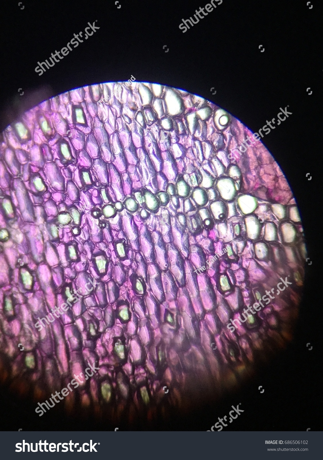
rhoeo discolor leaf cell, plants cell by Royalty Free Stock Photo 686506102
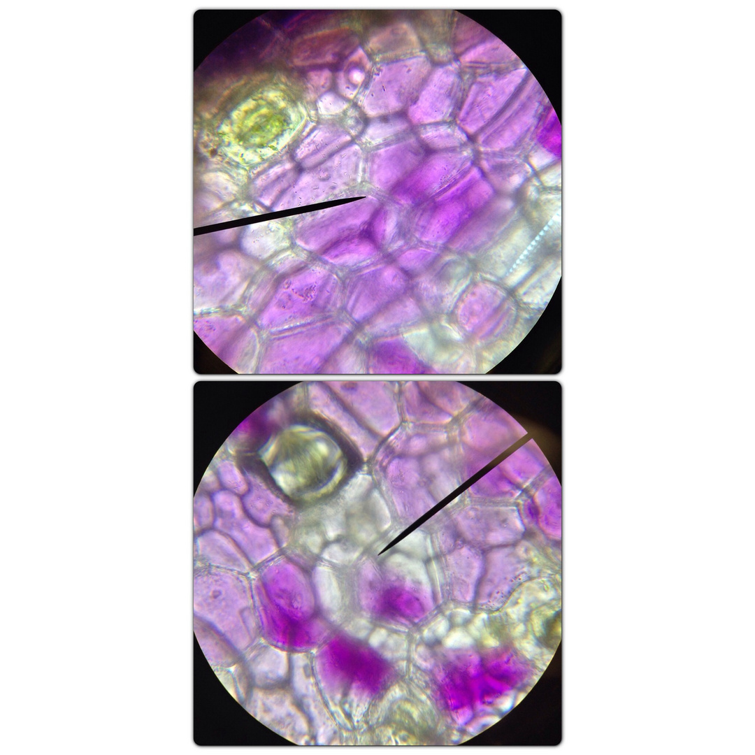
Diambil daun Rhoeo discolor, dibuat preparat sayatan daun seperti kegiatan 2. Diamati di bawah mikroskop dengan perbesaran 10 x 10. Diperhatikan stomata dan trikomatanya. Digambar bagaimana bentuk sel penutupnya! Jika kurang jelas maka digunakan perbesaran 10 x 45. Digambar dan diberi keterangan!

Rhoeo Discolor Cells with Plasmolysis Use Miksscope Stock Photo Image of plasmolysis
Tabel 3. 1 Alat dan Bahan Praktikum Alat Bahan Mikroskop Air Kaca objek Alkohol 95% Pinset Kertas saring atau tissue Brix refraktometer Bunga Rhoeo discolor Tabung reaksi Daun Hydrilla verticillata Umbi kentang Larutan I2KI Daun Ficus elastica Batang suji (Pleomele angustifolia) Tangkai daun Carica papaya Larutan cuka Tempurung kelapa Anilin.

Rhoeo Discolor Leaf (W.M) under the Microscope 🌿🔬 PeakD
Size. Rhoeo discolor is a compact plant, typically reaching heights of 6 to 12 inches (15 to 30 cm) with a similar spread.It has a moderate growth rate, with an average monthly growth of about 1 inch (2.5 cm) under optimal conditions.The plant produces a dense cluster of roots that are fibrous and relatively shallow, which allows it to be easily grown in pots or confined spaces.

aluna_aishiteru OSMOSIS CAIRAN SEL pada RHOEO DISCOLOR
A healthy Rhoeo leaf from a potted plant is taken. A part of the peel was removed with the help of forceps from the lower surface of the leaf. The peel was then placed in a watch glass, which contain water in it. With the help of forceps the peel was placed on a clean glass slide. Lastly the slide was observed under the microscope.

🧬🔬🧪 PRAKTIKUM DAN PENGAMATAN PERISTIWA OSMOSIS PADA RHOEO DISCOLOR DENGAN MENGGUNAKAN MIKROSKOP
Learn how to observe the plasma flow and the ergastic substances in various plants, such as Rhoeo discolor, Vallesneria, Solanum tuberosum, Ficus elastica, Pleomele angustifolia, and Carica papaya.

FileRhoeo Discolor epidermis.jpg Wikimedia Commons
Traditional medicine has led to the discovery of important active substances used in several health-related areas. Phytochemicals in Rhoeo discolor extracts have proven to have important antimicrobial activity. In the present study, our group determined the antimicrobial effects of extracts of Rhoeo discolor, a plant commonly used in Mexico for both medicinal and ornamental purposes.

difusi&osmosis, plasmolisis dan tekanan turgor Prapatan SPS
Studi Potensi Ekstrak Daun Adam Ha wa (Rhoeo discolor) Sebagai Indikator Titras i Asam- Basa Ratnasari, S., Suhendar, D., Amalia, V. 485 505 525 545 565 585 605 625

Rhoeo discolor up close and personal Leaf images, Galaxy art, Nature inspiration
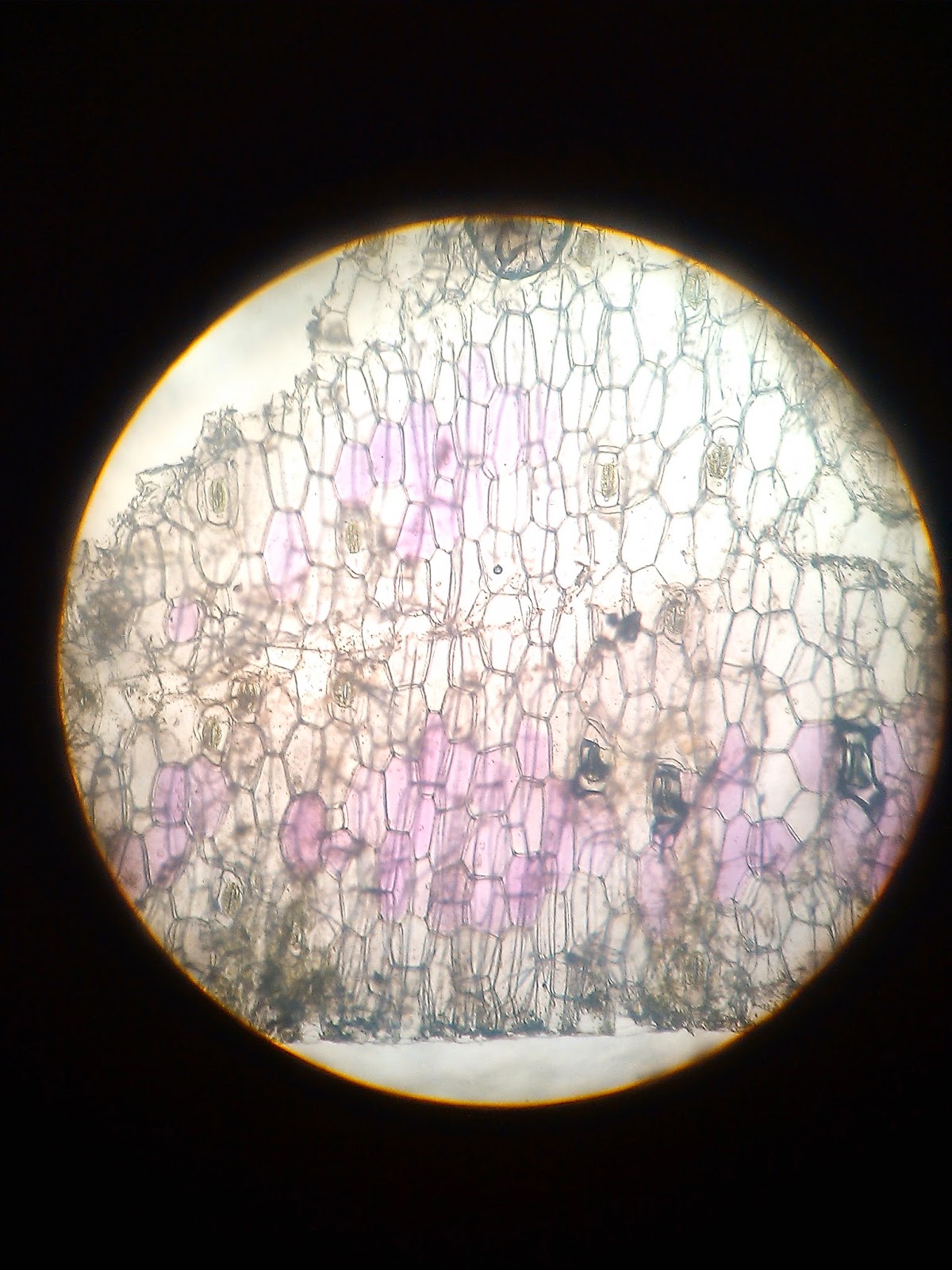
Setelah membuat preparat dari irisan daun adam hawa (Rhoeo discolor) dan mengamatinya di bawah mikroskop dengan perbesaran 15 x 10, terlihat struktur jaringan daun adam hawa yang terdiri dari dinding sel yang merupakan bagian luar dari masing- masing sel, sitoplasma yang merupakan suatu suspensi yang berada di dalam sel, stomata yang merupakan alat pernapasan serta sel penjaga yang berfungsi.
Gambar Sel Rhoeo Discolor bonus
Traditional medicine has led to the discovery of important active substances used in several health-related areas. Phytochemicals in Rhoeo discolor extracts have proven to have important antimicrobial activity. In the present study, our group determined the antimicrobial effects of extracts of Rhoeo discolor, a plant commonly used in Mexico for both medicinal and ornamental purposes. We.

Yuk, Ketahui Terjadinya Plasmolisis pada Daun Rhoeo Discolor
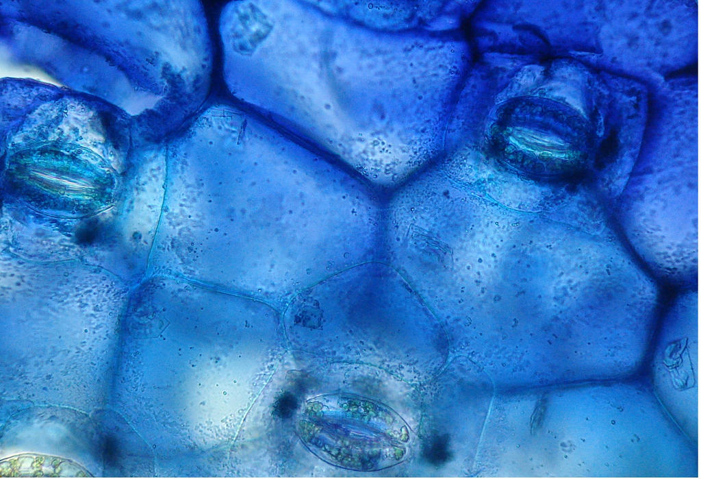
Pada pengamatan daun Rhoeo discolor pada mikroskop cahaya terlihat jaringan epidermis pada daun Rhoe discolor yang berbentuk persegi panjang dan susunan selnya rapat yang berfungsi sebagai pelindung sel-sel yang ada dibawahnya. Dan juga terdapat stoma,stoma ini terdiri dari satu porus atau celah dan dua sel penutup yang mengapitnya.

Anatomi Melintang Daun Rhoeo discolor YouTube
Assalamualaikum wr wb… Haloo PECINTA BIOLOGI! Sudahkah kalian memahami cara membuat preparat dan mengamati preparat Rhoeo Discolor dengan mikroskop? Yuk bela.
Rhoeo discolor skin cells by Etherbreeze on DeviantArt
Corresponding Author. Rhoeo discolor is a plant used in traditional medicine mainly due to its anticancer properties. The present work studied, for the first time, its use as a phytoremediation plant. Samples of R. discolor were collected in the gardens of the Universidad Ju?rez Aut?noma de Tabasco.

aluna_aishiteru OSMOSIS CAIRAN SEL pada RHOEO DISCOLOR
Minggu, 15 Maret 2020. perbesaran 10 x 10 mikroskop; daun Rhoeo discolor. LAPORAN PRAKTIKUM DAUN RHOEO DISCOLOR (Nanas Kerang) A. Tujuan : 1. Siswa diharapkan mampu untuk mengetahui struktur dan bentuk jaringan tumbuhan. 2. Siswa mampu mengetahui bentuk stomata daun Rhoeo Discolor (Nanas Kerang) B. Landasan Teori.

Anatomi Membujur Daun Rhoeo discolor YouTube
This study was focused on students visual spatial ability in representing single cell epidermis of Rhoeo discolor leaf. It was conducted with 44 undergraduate students who presently enchanting.

Detail Gambar Sel Daun Rhoeo Discolor Koleksi Nomer 23
Plant material. The typical ring-forming (ring of 12 chromosomes) variety known also as Rhoeo discolor (Golczyk et al. 2005, 2010) and the bivalent-forming (6 bivalents) variety concolor obtained from Kew Botanical Garden (Golczyk 2011c) were grown in pots filled with soil in a greenhouse at 25-27 °C.Their young ca. 2-4-mm-long flower buds and small leaf fragments were excised and fixed.
Plasmolyse bei Rhoeo discolor vorhernachher Foto & Bild sonstiges, fossilien & präparate
Daun Rhoeo discolor 2. Mikroskop 3. Larutan Gula 5% 4. Aquades 5. Gilet ( Silet ) 6. Gelas Objek 7. Cover Glass 8. Kertas Isap 9. Pipet 10. Kamera C. Cara Kerja C.A Pengamatan daun Rhoeo discolor dengan menggunakan air 1. Menyiapkan daun Rhoeo discolor, kemudian menyanyatnya secara membujur. 2.