
Maxillary First Premolar Anatomy ANATOMY STRUCTURE
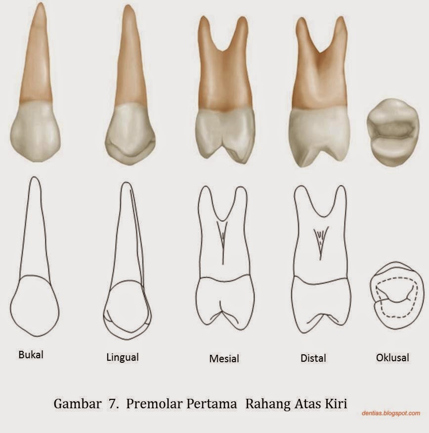
2. Gigi Premolar Di belakang gigi kaninus ada gigi premolar. Bentuk gigi premolar di rahang atas agak berbeda dengan premolar di rahang bawah. Premolar rahang atas mempunyai dua bonjol, sedangkan premolar rahang bawah hampir mirip dengan kaninus namun bonjolnya tidak runcing dan bentuknya juga lebih besar dari gigi kaninus.

Arch traits that differentiate maxillary from mandibular premolars Pocket Dentistry
The premolar teeth (Latin: dentes premolares), also known as premolars and bicuspids, lie between the canines and first molar teeth.. The oral cavity houses four maxillary premolars (4, 5, 12, 13) in the upper jaw and four mandibular premolars (20, 21, 28, 29) in the lower jaw.Each side of the jaw (quadrant) contains two premolars. Overall, every adult has 8 premolar teeth.
The Permanent Maxillary Premolars (Dental Anatomy, Physiology and Occlusion) Part 1
Human beings typically have eight total premolars. There are two premolars in each of the four dental quadrants of the mouth. Although the exact age at the time of arrival varies, the first premolar usually arrives around the age of 10-11 years old and the second premolar usually comes in around the age of 10-12 years old.
Jual Gigi premolar rahang bawah Shopee Indonesia
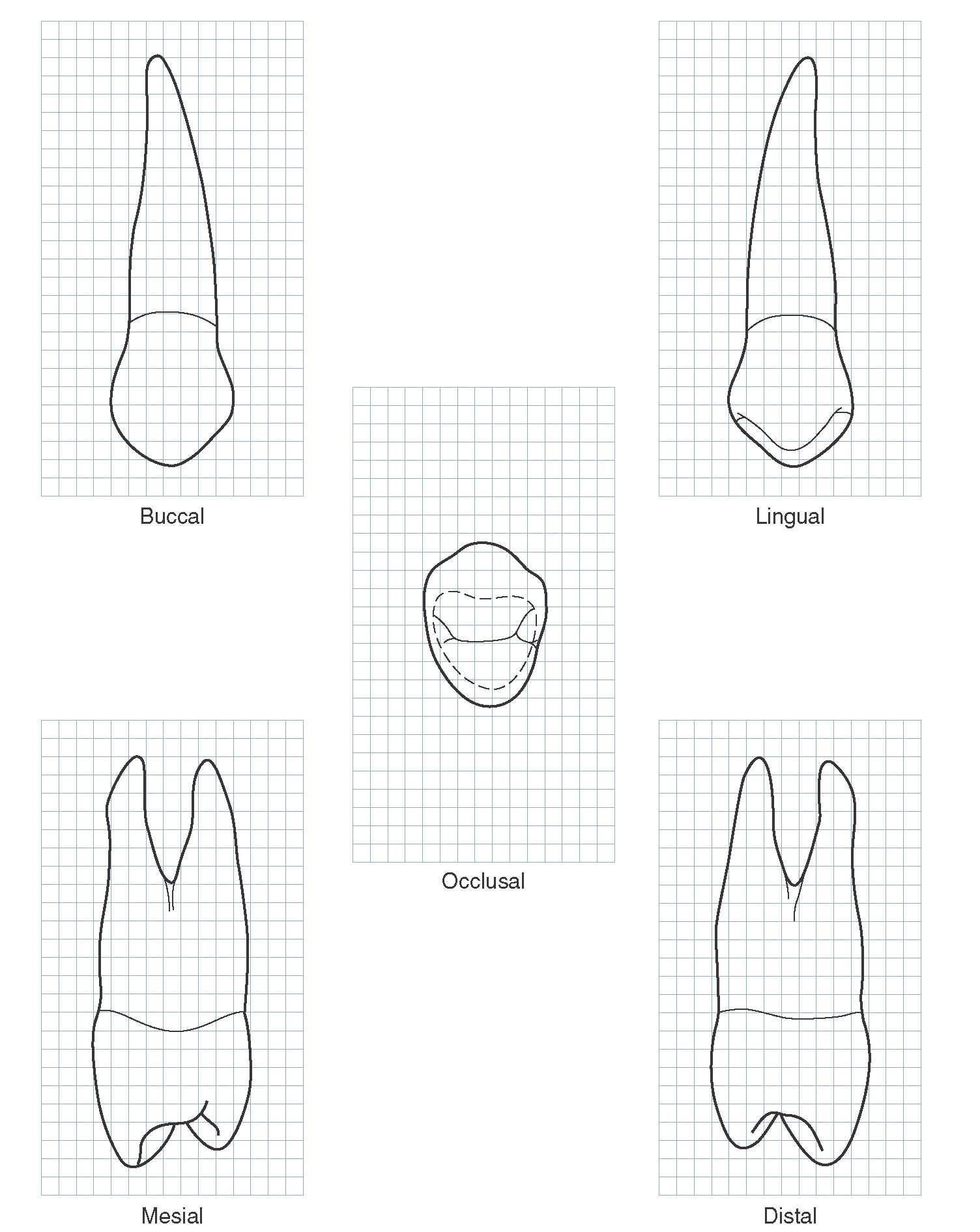
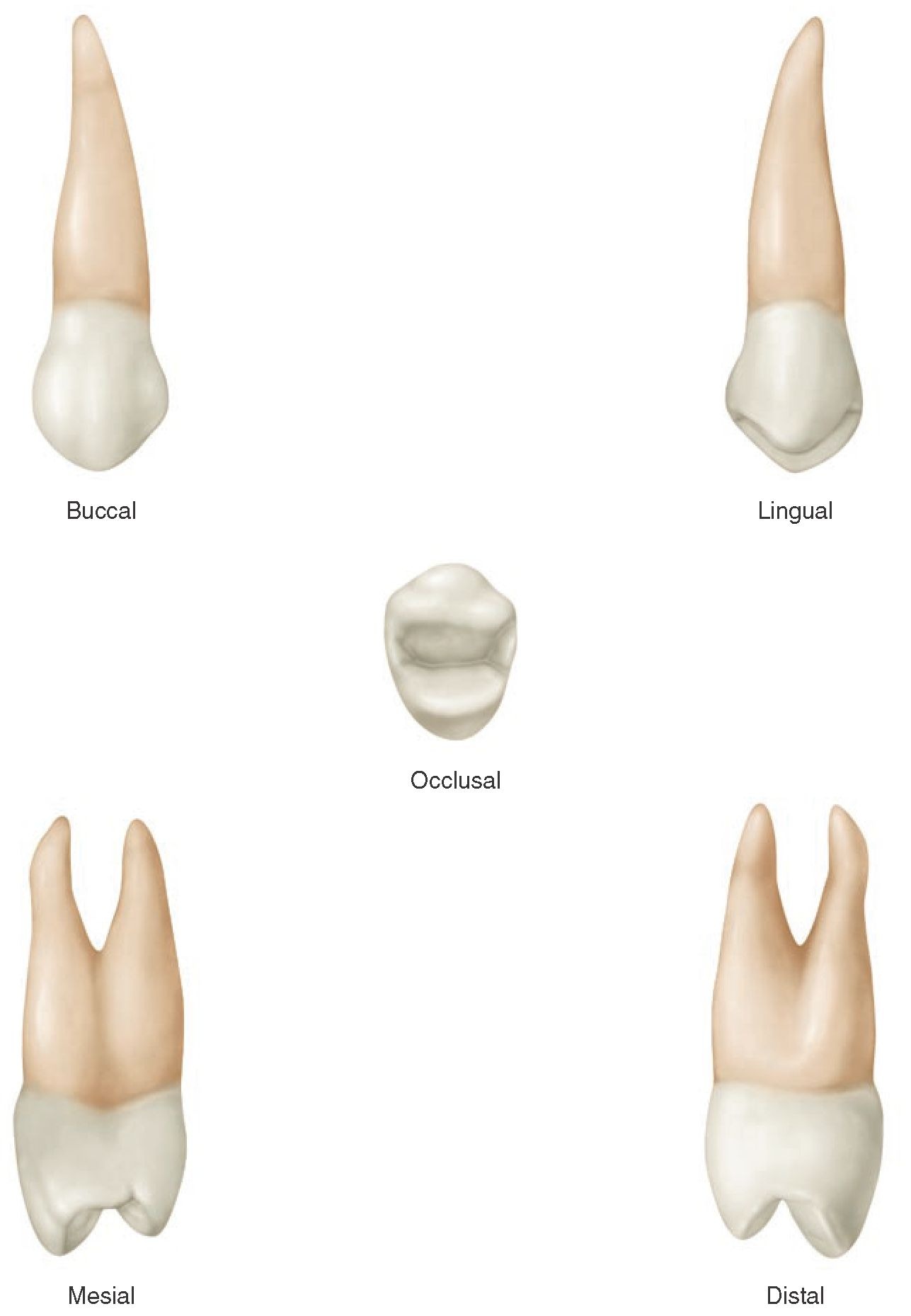
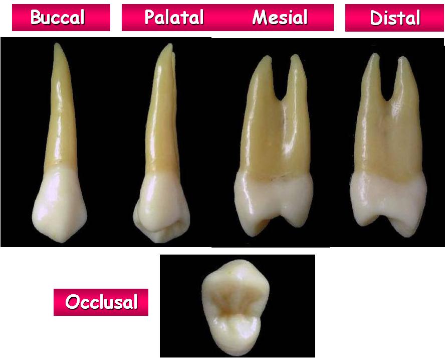
Maxillary First Premolar. Figures 9-1 through 9-16 illustrate the maxillary first premolar from all aspects. The maxillary first premolar has two cusps, a buccal and a lingual, each being sharply defined. The buccal cusp is usually about 1 mm longer than the lingual cusp. The crown is angular, and the buccal line angles are prominent.
PPT Premolars PowerPoint Presentation, free download ID4246791
Gigi Molar Satu Rahang Atas dan Bawah di Jawa Barat Penelitian ini bertujuan untuk menginvestigasi jumlah dan bentuk akar serta konfigurasi saluran akar pada gigi molar satu atas dan bawah di Jawa Barat, Indonesia. 100 molar satu atas dan 100 molar satu bawah bawah dikumpulkan dari praktek dokter gigi. Dilakukan perhitungan jumlah akar dan derajat

The Root Canal Anatomy Project Mandibular First Premolar
Gigi geraham kecil ke-1 atau premolar 1 (usia 10-11 tahun) Gigi geraham kecil ke-3 atau premolar 2 rahang atas dan rahang bawah (usia 10-12 tahun) Gigi taring (usia 11-12 tahun) Gigi geraham ke-2 (usia 12-13 tahun) Faktor yang Memengaruhi Pertumbuhan Gigi Permanen Tidak semua anak mengalami pertumbuhan gigi permanen sesuai waktunya.
The Permanent Maxillary Premolars (Dental Anatomy, Physiology and Occlusion) Part 1
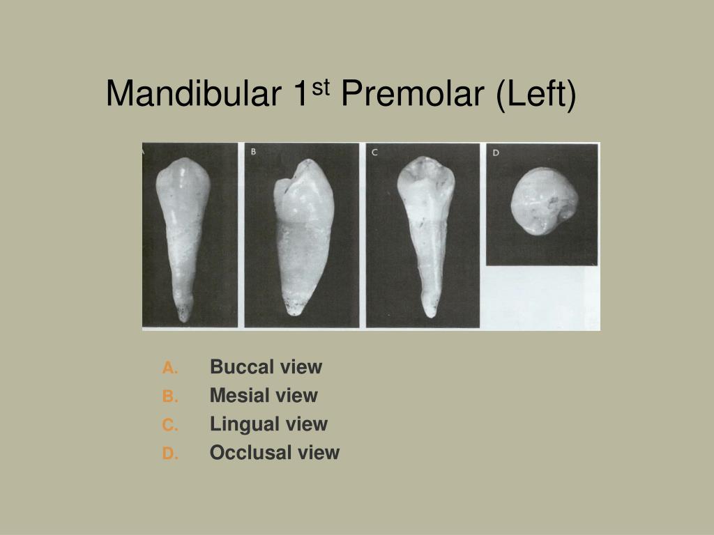
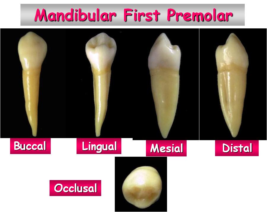
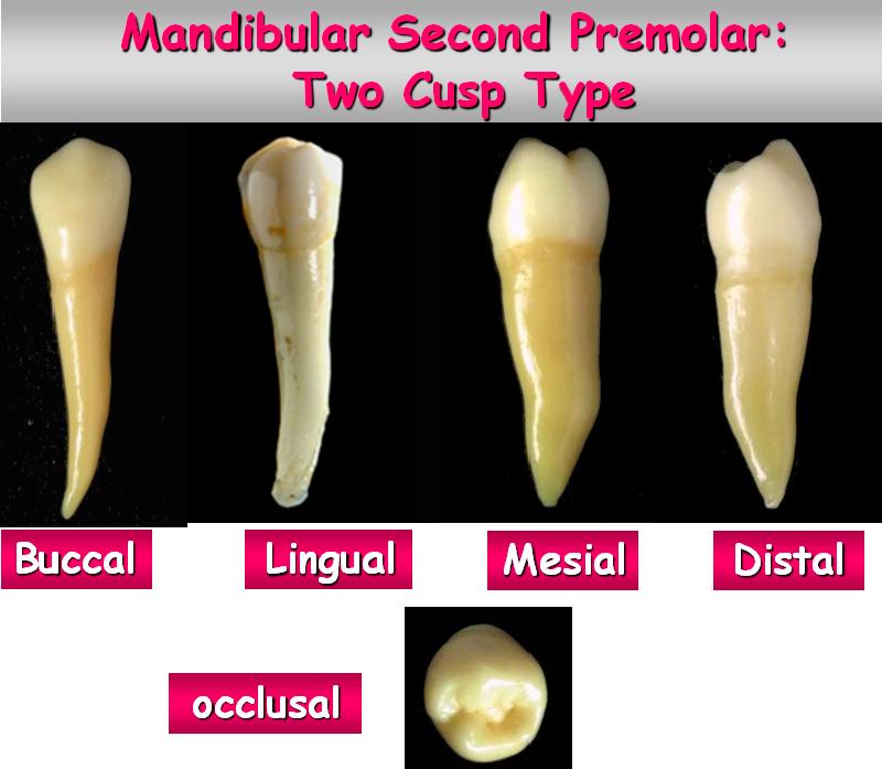
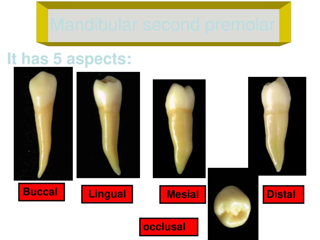
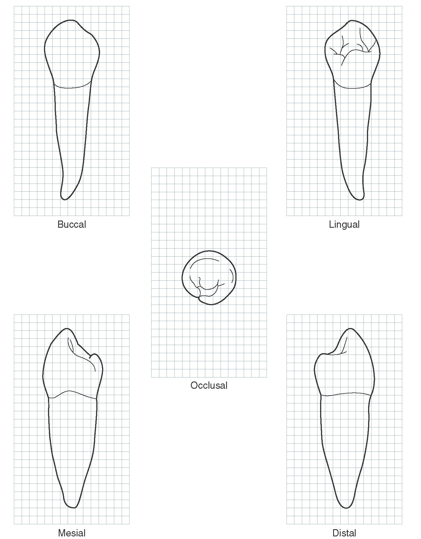
PREMOLAR 1 BAWAH • Gigi ke 4 dari median line rahang bawah • Tugas sama dg caninus dan P 1 atas2 • 2 cusp, cusp mirip dengan caninus dan lebih panjang bukal cusp • Akar 1, bundar dan meruncing, membelok kedistal • Groove developmental nyata sekali • Bentuk perm oklusal bulat
RxDentistry DENTAL ANATOMY OF PREMOLARS
The first mandibular premolar presents with two cusps- buc cal and lingual.The buccal cusp is very large, while the lingual cusp is very small. This characteristic allows seeing the entire occlusal surface when looking at it from the lingual surface of the tooth.
Morfologi Gigi Permanen Dentias notes
Ringkasan Apa itu Premolar Pertama? Premolar pertama adalah salah satu dari premolar yang diberi nama berdasarkan distribusinya. Dengan demikian, premolar pertama dapat diklasifikasikan sebagai premolar mandibula pertama dan premolar rahang atas pertama. Premolar rahang bawah pertama terletak lateral di rahang bawah jauh dari garis tengah wajah.

one visit endodontic treatment maxillary premolar vital pulp one curve micromega YouTube
Similar to 10. morfologi gigi permanent rahang bawah (11) Morphology of Permanent Teeth. Primary teeth original. Determinasi gigi molar decidui bawah syiiiik. Determinasi gigi molar decidui bawah syiiiik. Premolar pertama rahang atas. 2. anatomi gigi insisivus sentral bawah kanan. Anatomi gigi permanen premolar. Premolar kedua rahang atas.
RxDentistry DENTAL ANATOMY OF PREMOLARS
Laporan kasus Perawatan saluran akar (PSA) satu kali kunjungan pada gigi molar pertama bawah kanan dengan restorasi endocrown resin komposit
Dentistry lectures for MFDS/MJDF/NBDE/ORE A Note on Dental Anatomy of Premolars
Premolar kedua bawah Gigi ini adalah gigi ke-5 dari garis median. Meskipun ukuran mesio-distal dari korona dan akar hampir sama seperti premolar pertama bawah,tetapi pertumbuhannya pada lain permukaan lebih baik Pada gigi ini terdapat 2 jenis yaitu: 1. Premolar dengan 3 cusp(1 cusp bukal dan 2 cusp lingual) 2.
PPT Mandibular first premolar PowerPoint Presentation, free download ID4341166
premolar 1 rahang atas dan rahang bawah dalam . hal kesesuaian hasil penilaian usia dentalis dengan . usia kronologis yang me nunjukkan bahwa usia . dentalis sangat mendeka usia kronologis pada saat .
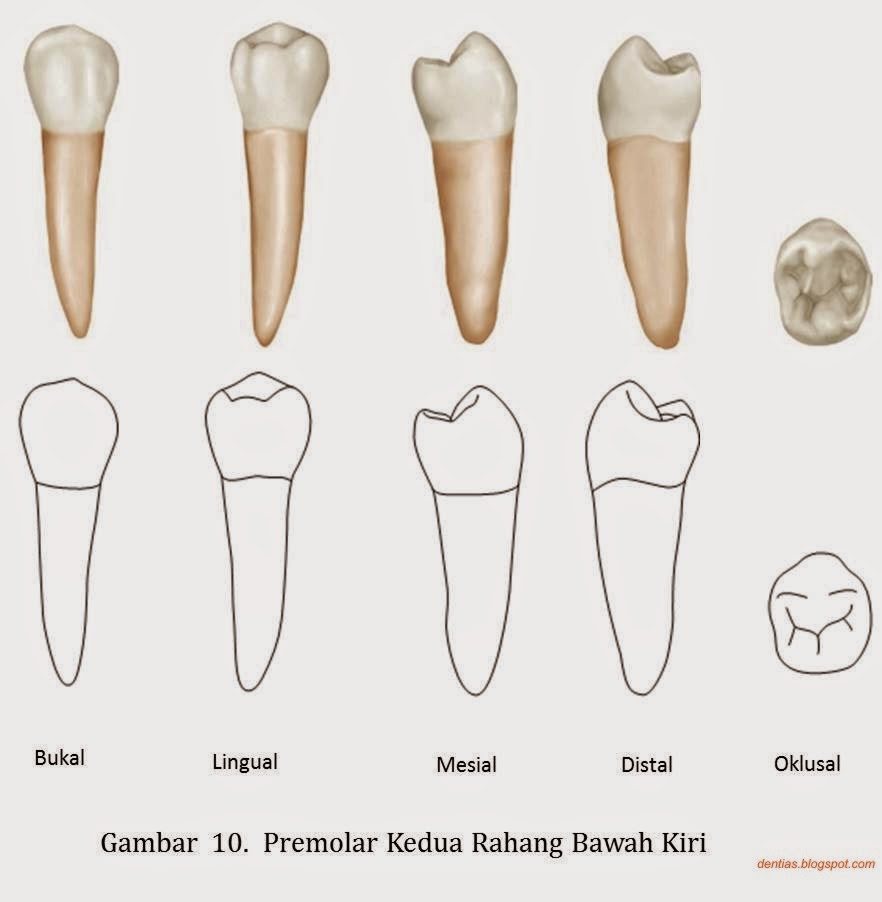
Morfologi Gigi Permanen Dentias notes
Penjelas Gambar Anatomi Gigi, Jenis Gigi, dan Fungsi Bagiannya Gigi adalah bagian dalam tubuh manusia yang cukup rumit. Fungsi gigi tidak hanya untuk mengunyah dan mencerna makanan, tapi juga berperan penting dalam berbicara. Untuk mengetahui lebih dalam tentang gigi, simak anatomi gigi selengkapnya di sini. Mengenal perkembangan struktur gigi

6. anatomi gigi premolar 1 & 2 bawah
The premolars, also called premolar teeth, or bicuspids, are transitional teeth located between the canine and molar teeth. In humans, there are two premolars per quadrant in the permanent set of teeth, making eight premolars total in the mouth. [1] [2] [3] They have at least two cusps.
The Permanent Mandibular Premolars (Dental Anatomy, Physiology and Occlusion) Part 1
The first maxillary premolar contains two cusps called the buccal and lingual cusps.The lingual cusp is shorter and smaller, and it is slightly displaced to the mesial surface.The highest point of each cusp is called the cusp apex. Therefore, the first maxillary premolar presents with the buccal and lingual cusp apexes.. The buccal surface presents two developmental grooves (depressions) - the.