
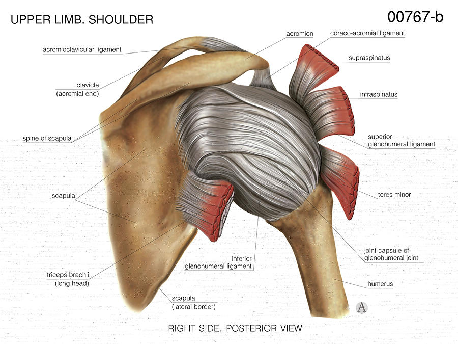
079 The Ligaments of the Glenohumeral Joint Interactive Biology, with Leslie Samuel
Introduction. Subacromial impingement syndrome (SIS) is a clinical syndrome most often attributed to patients presenting with shoulder pain. 1 SIS is a syndrome that encompasses a spectrum of subacromial pathologies ranging from bursitis, rotator cuff tendinosis, and partial tears leading up to full-thickness tear of the rotator cuff. Luime et al. estimated the prevalence of shoulder.
Shoulder Replacement New Mexico Orthopaedic Associates
Latar Belakang : Menurut Lampignano dan Kendrick (2018) proyeksi, yang digunakan pada pemeriksaan radiografi shoulder joint pada pasien trauma yaitu Anterior posterior (Neutral Rotation), Trans thoracic Lateral, PA Oblique Scapular Y lateral), Proyeksi Tangensial (Supraspinatus Outlet), AP Apical Oblique. Menurut Standar Prosedur Operasional (SPO) di RSUD RAA Soewondo Pati prosedur pemeriksaan.

Mr Paul Jarrett Shoulder Anatomy Murdoch Orthopaedic Clinic
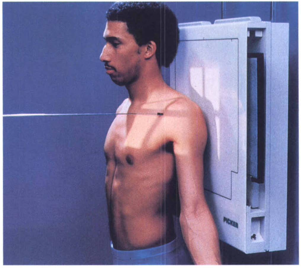
Teknik pemeriksaan shoulder joint metode . scapular "Y" dengan posisi pasien berdiri tegak, posisi objek menghadap ke IR dan bagian yang . diperiksa menempel pada IR. Menurut Frank, Long,

Shoulder joint, MRI scan Stock Image C031/5282 Science Photo Library
Teknik pemeriksaan radiografi shoulder joint proyeksi inferosuperior merupakan pemer iksaan shoulder joint dengan posisi pa sien supine dan l engan di abduksi sebesar 90 o .
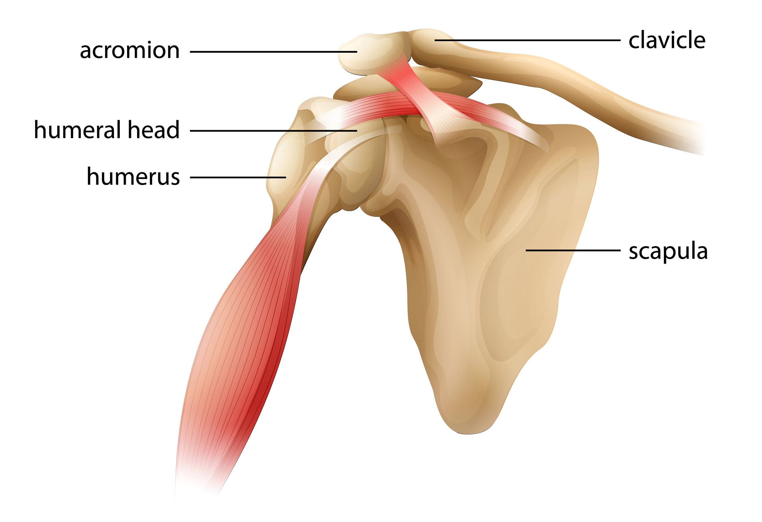
Shoulder Joint Anatomy
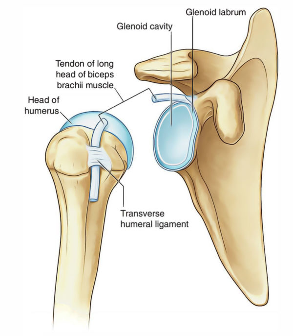
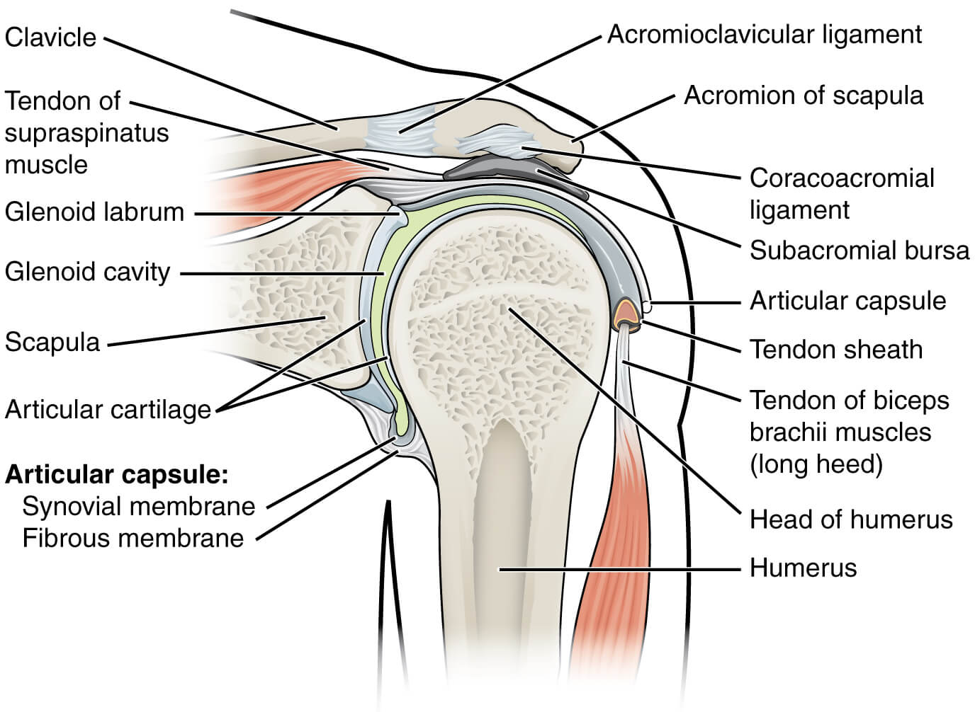
Shoulder joint adalah ball-and-socket joint yang terbentuk oleh head humerus dan glenoid cavity dari tulang scapula. Dislokasi sendi merupakan keadaan di mana tulang- tulang yang membentuk sendi tidak lagi berhubungan secara anatomis. proyeksi yang digunakan dalam kasus dislokasi ini yaitu AP dan Lateral.Tujuan penelitian ini yaitu mengetahui teknik radiografi pada pemeriksaan shoulder joint.
Shoulder Joint (Glenohumeral Joint) Earth's Lab
shoulder joint motion. Tendinitis in M.Rotator Cuff, is an inflammation occured in muscle tendons fused in Rotator Cuff. Physiotherapy problematics of Frozen Shoulder are the existence of. Pemeriksaan yang dilakukan yaitu pemeriksaan nyeri, pemeriksaan keterbatasan lingkup gerak sendi, pemeriksaan kekuatan

LOMBA VIDEOGRAFI TEKNIK PEMERIKSAAN CLAVICULA DAN SHOULDER JOINT YouTube
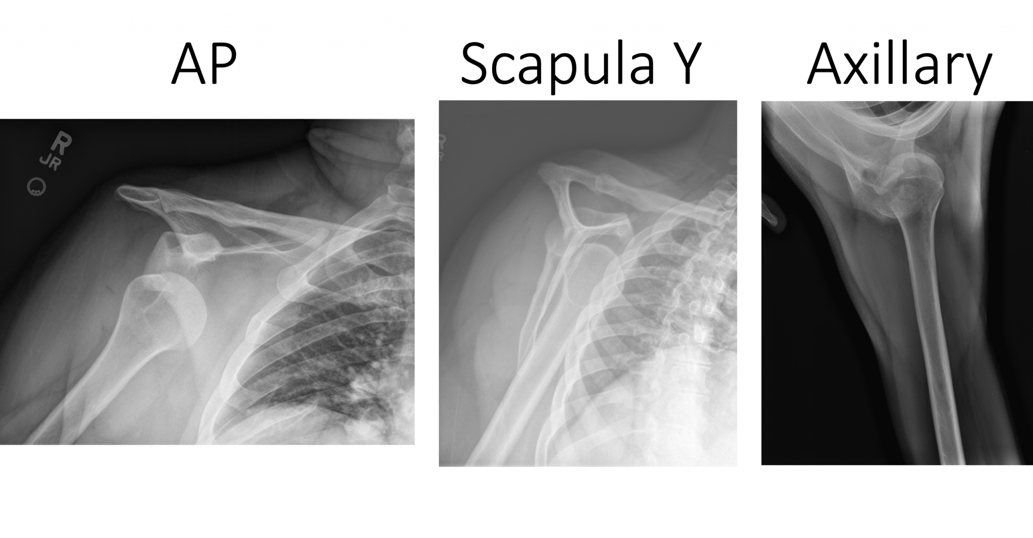
Hasil penelitian menunjukkan proyeksi pemeriksaan yang optimal pada pemeriksaan shoulder joint pada kasus trauma bahu adalah Anteroposterior (AP) dikarenakan menampakkan anatomi shoulder joint secara keseluruhan sehingga dapat mendiagnosa apabila terdapat dislokasi atau fraktur kemudian Scapular Y View sebagai proyeksi tambahan untuk.
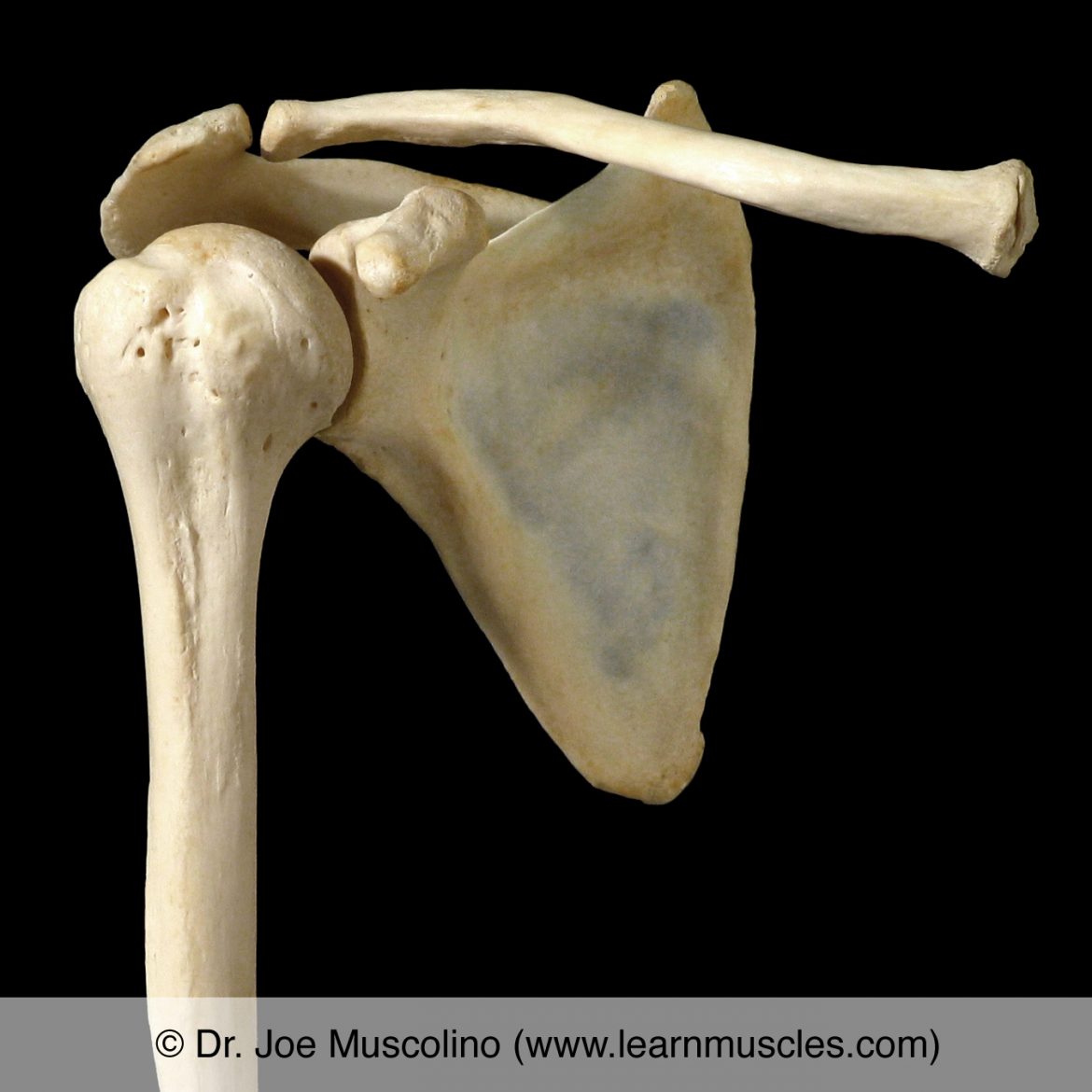
Shoulder (Glenohumeral) Joint Learn Muscles
Pemeriksaan shoulder joint adalah teknik pemeriksaan menggunakan sinar x untuk melihat struktua anatomi dari sendi bahu. Pemeriksaan ini dilakukan dengan beberapa proyeksi, yaitu proyeksi Antero Posterior (AP) external, neutral dan internal rotation humerus. transthoracic lateral, Antero Posterior (AP) axial, Antero Posterior (AP) oblique.

Shoulder Joint AnatomySkeletal SystemCartilagesLigamentsMusclesTendons
pemeriksaan MRI shoulder joint dan sampel pada penelitian ini 10 orang sukarelawan. Prosedur penelitian ini adalah Pemeriksaan dilakukan dengan sekuen Gradient Echo T2* menggunakan posisi netral
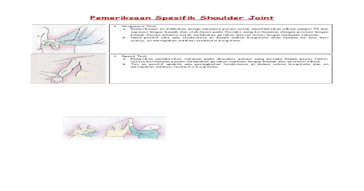
Pemeriksaan Spesifik Shoulder Joint [PDF Document]
The prerequisite for any treatment in the shoulder region of a patient with pain is a precise and comprehensive picture of the signs and symptoms as they occur during the assessment and as they existed until then. Because of its many structures (most of which are in a small area), its many movements, and the many lesions that may occur either inside or outside the joints, the shoulder complex.

Healthy shoulder joint, MRI scan Stock Image C048/9209 Science Photo Library
Dr. Ebraheim's educational animated video describes clinical evaluation tests of the shoulder.Follow me on twitter:https://twitter.com/#!/DrEbraheim_UTMCFind.
Shoulder Joint Pain Causes & Treatment Dr. Chris Homan
Traditionally Orthopaedic Special tests were used to assist in the diagnostic process by implicating specific tissue structures that are either dysfunctional, pathological, or lack structural integrity, confirming the findings from the physical assessment and providing a tentative diagnosis. [1] Special testing is generally performed following.
Gudang Medis Teknik Radiografi Shoulder Joint
Teknik Pemeriksaan Radiografi Shoulder. Dipos oleh Rini pada Juni 13, 2021. Proyeksi : AP (External Rotation) Kaset : ukuran 24 x 30 cm. kV : 70 ± 5 mAs : 6. FFD : 100 cm. Posisi Pasien : Posisi pasien erect atau supine. (Posisi erect biasanya mengurangi sakit pada pasien) Posisi Obyek : Putar tubuh ke shoulder yang sakit sehingga menempel ke.
Acromioclavicular Joint Separation Undergraduate Diagnostic Imaging Fundamentals
Studi literatur ini bertujuan untuk Mengetahui prosedur teknik pemeriksaan radiografi shoulder joint pada kasus Dislokasi menurut Timothy G. Sanders (2005), Bruno and Patrick (2016), Scott E. Sheehan (2012). Jenis penelitian ini adalah kualitatif dengan pendekatan studi literatur,waktu penelitian oktober2020-Maret2021. Pengumpulan data.
Emergency Medicine EducationEM3AM Anterior Shoulder Dislocation
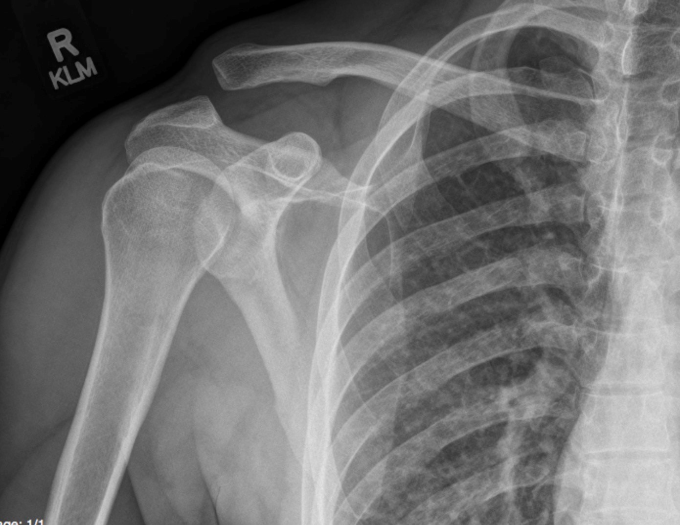
Citation, DOI, disclosures and article data. The shoulder AP view is a standard projection that makes up the two view shoulder series. The projection demonstrates the shoulder in its natural anatomical position allowing for adequate radiographic examination of the entire clavicle and scapula, as well as the glenohumeral, acromioclavicular and.
Glenohumeral Joint WikiMSK
The shoulder joint dislocates more frequently than any other joint in the body. A dislocation can become worse by strained or torn fibrous tissue which connects the bones. The bones can only be pulled out of position by a powerful force, such as a blow to the shoulder. Extreme rotation can cause the humeral head to come out of the glenoid labrum.