Intervertebral Disc Anatomy
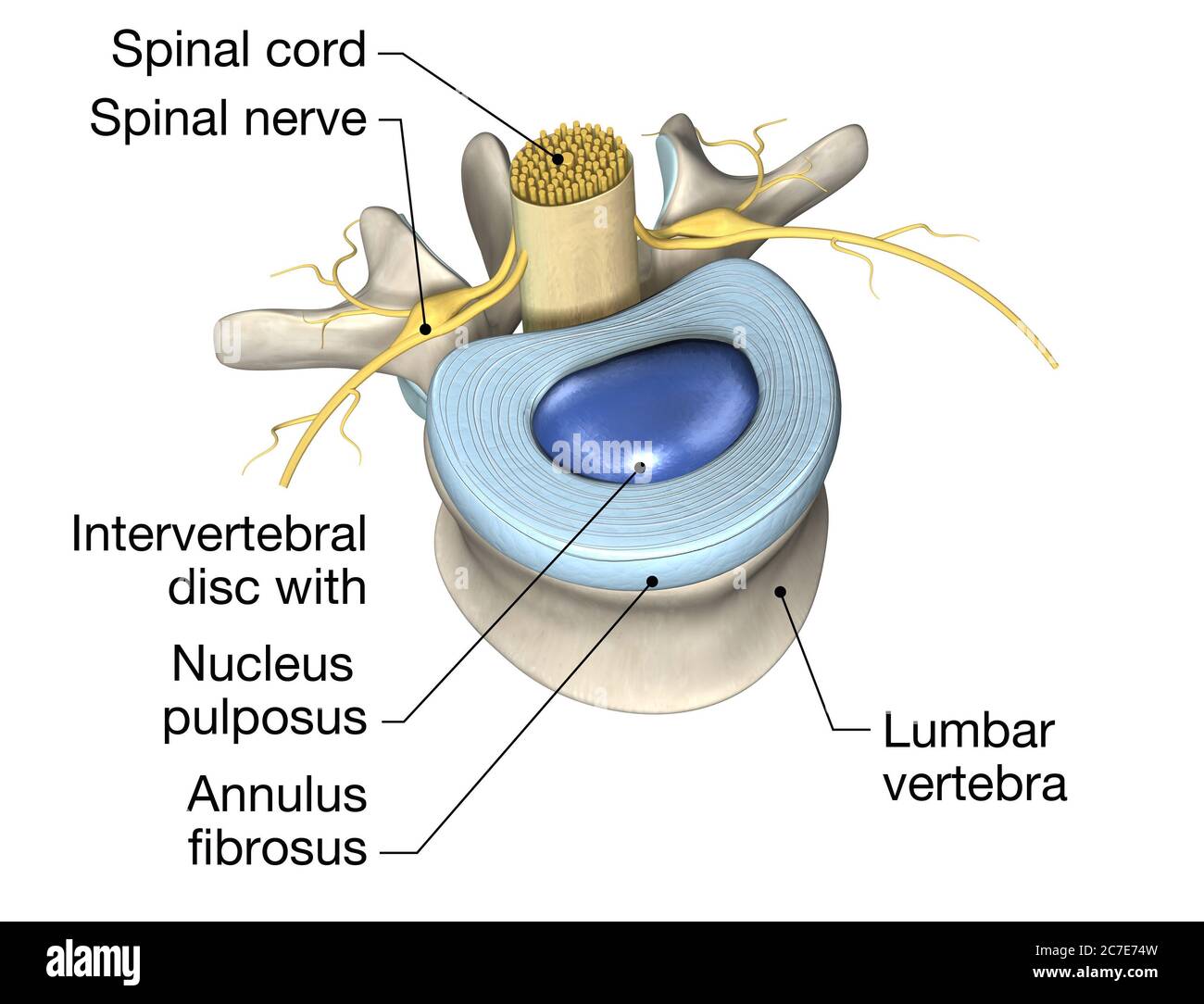
The intervertebral discs are secondary cartilaginous joints, also known as symphyses 7 . Each intervertebral disc is comprised of: peripheral annulus fibrosus. central nucleus pulposus. hyaline cartilage (vertebral side) and fibrocartilage (nucleus pulposus side) Above and below the intervertebral disc are the vertebral body endplates .
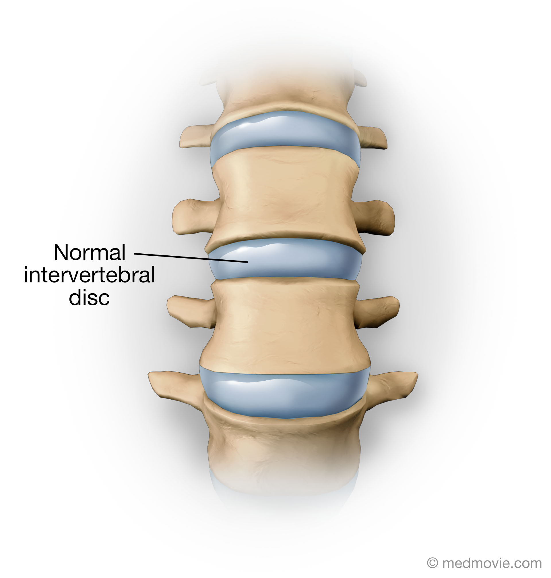
Normal Intervertebral Disc
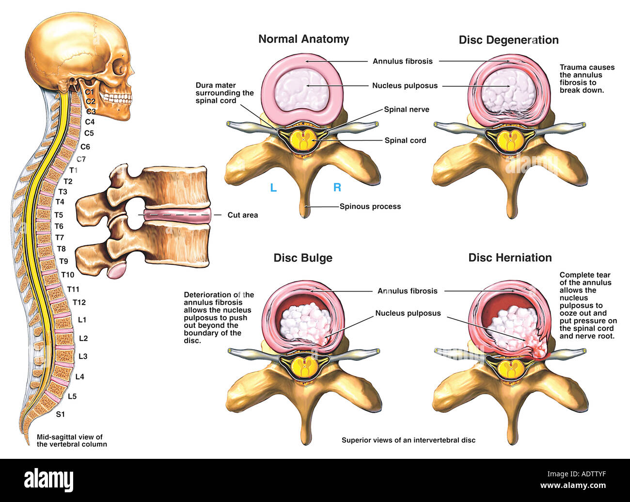
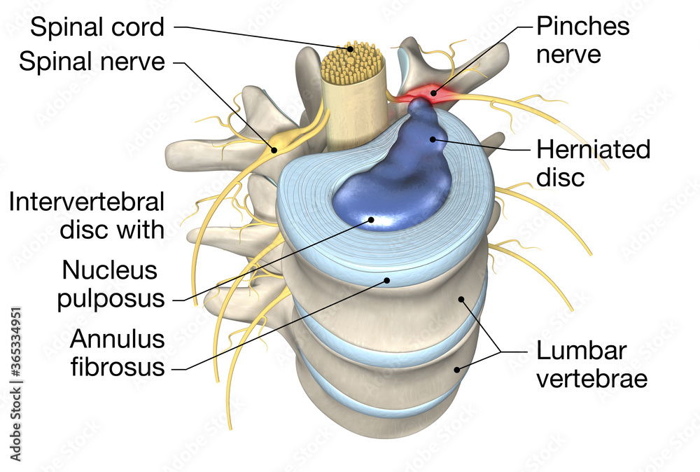
A spinal disc herniation is an injury to the intervertebral disc between two spinal vertebrae, usually caused by excessive strain or trauma to the spine. It may result in back pain, pain or sensation in different parts of the body, and physical disability. The most conclusive diagnostic tool for disc herniation is MRI, and treatment may range.
Intervertebral disc T12L1 (Discus intervertebralis TXIILI) Kenhub
Intervertebral discs consist of an outer fibrous ring, the anulus (or annulus) fibrosus disci intervertebralis, which surrounds an inner gel-like center, the nucleus pulposus. The anulus fibrosus consists of several layers (laminae) of fibrocartilage made up of both type I and type II collagen.Type I is concentrated toward the edge of the ring, where it provides greater strength.
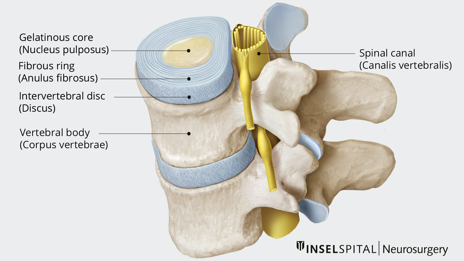
Herniated Disc Neurosurgery Inselspital Bern
Diskus Intervertebralis adalah cakram yang membentuk sendi tulang rawan di antara vertebra, memberikan peredam kejut yang sangat efisien. Diskus intervertebralis terdiri dari anulus fibrosus, nukleus pulposus dan tulang rawan di ujung lempengnya. Struktur ini sering rusak dan menjadi penyebab paling umum gangguan tulang punggung bawah.
Intervertebral Discs Anatomy and Embryology Kenhub
Citation, DOI, disclosures and article data. Vacuum phenomena involving the intervertebral discs is usually a result of an accumulation of gas, principally nitrogen , within the crevices of the intervertebral discs or adjacent vertebrae. This is a joint-specific example of the vacuum phenomenon.

Diskus intervertebralis II Anatomi og Fysiologi
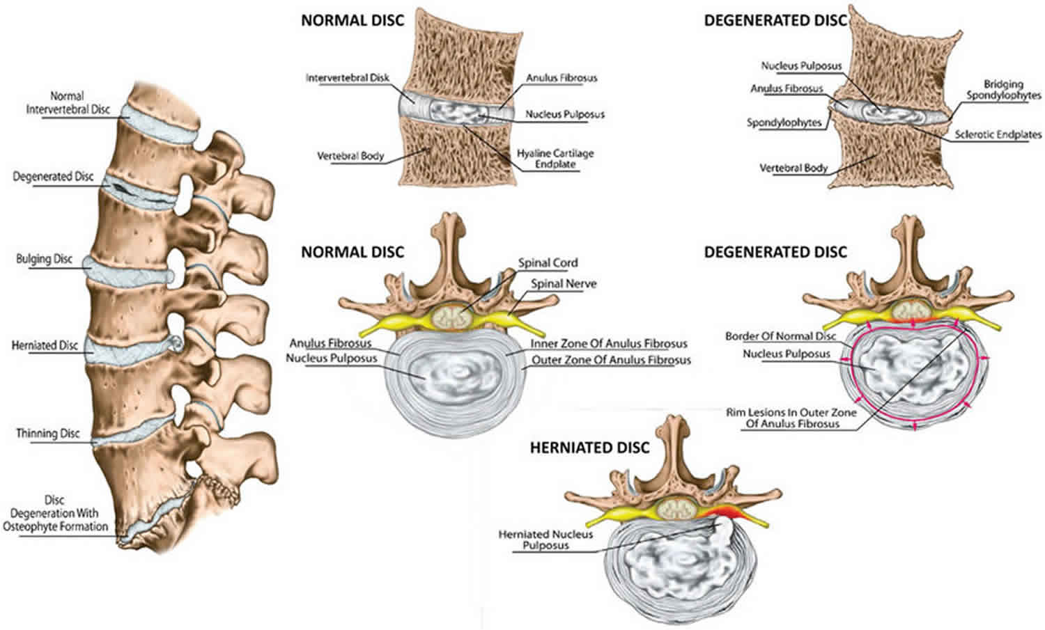
Intervertebral disc disease is a common condition characterized by the breakdown (degeneration) of one or more of the discs that separate the bones of the spine (vertebrae), causing pain in the back or neck and frequently in the legs and arms. The intervertebral discs provide cushioning between vertebrae and absorb pressure put on the spine.
Intervertebral disc Wikipedia
Diskus intervertebralis sebagian besar avaskuler atau tidak ada pembuluh darah. Struktur di diskus intervertebra mendapat nutrisi dan oksigen secara difusi dari pembuluh darah di sekitarnya. Seiring dengan bertambahnya usia seseorang maka organ dalam tubuh akan mengalami penuaan atau degenerasi, termasuk juga diskus intervertebra.
Intervertebral discs Anatomy and embryology Kenhub
Intervertebral disc degeneration (IVD) is characterized by a series of disruptive changes, structural and biochemical, which may lead to pain and disability. The appearance of these changes is usually irrespective of age, sex, and spinal level. The etiology of IVD degeneration is multifactorial, which can include genetic, environmental, altered.

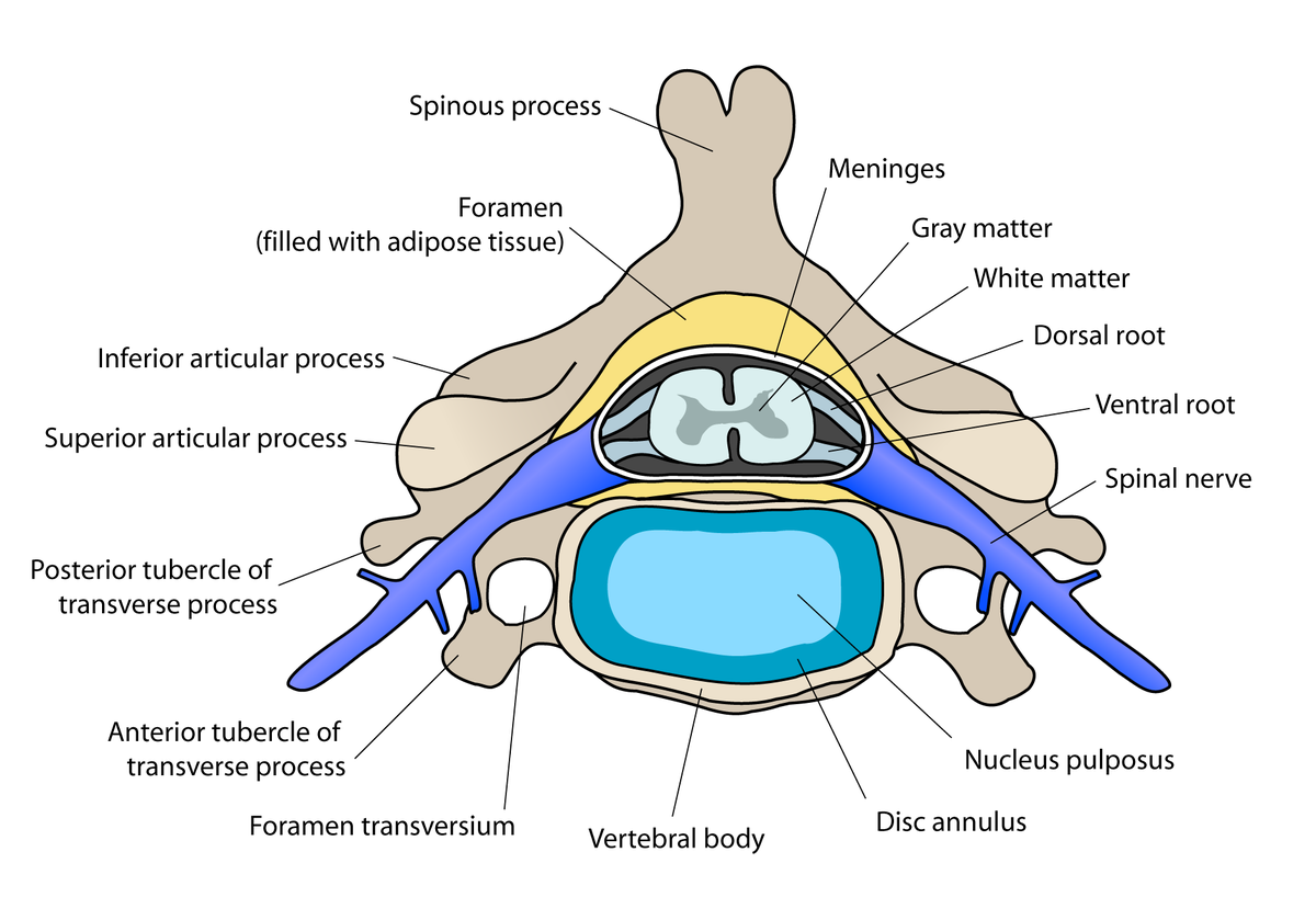
Anatomy Stock Images neckintervertebraldiscdiscusintervertebralisspinousprocessfacet
Jika dilakukan pada tulang belakang, istilah discus intervertebralis bisa saja muncul. Punggung kita terdiri dari banyak tulang belakang (vertebrae) yang tiap sela-selanya diisi oleh bantalan yang disebut discus intervertebralis. Tulang yang banyak dan bersela-sela ini penting untuk kita dapat melakukan gerakan membungkuk, membusung, dan.
Intervertebral Discs Anatomy and Embryology Kenhub
An intervertebral disc is a structure located between adjacent vertebrae of the spine. It consists of a tough outer layer called the annulus fibrosus and a gel-like center called the nucleus pulposus. The intervertebral disc acts as a cushion, allowing the spine to bend and twist without damaging the vertebrae.
3D illustration showing lumbal vertebra with intervertebral disc, medically 3D illustration
Discus intervertebralis. Definition. The intervertebral discs (intervertebral fibrocartilages) are interposed between the adjacent surfaces of the bodies of the vertebræ, from the axis to the sacrum, and form the chief bonds of connection between the vertebræ. They vary in shape, size, and thickness, in different parts of the vertebral column
Biology Intervertebral disc
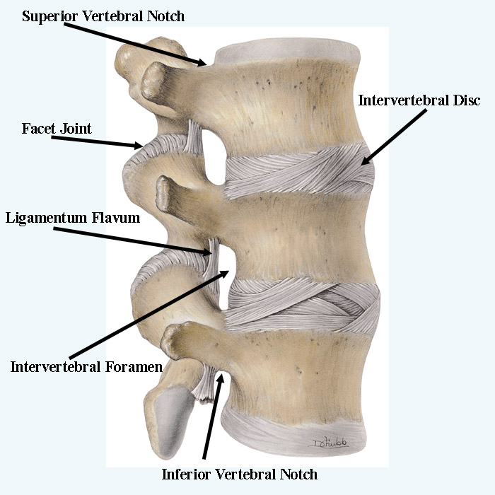
Synonyms: none. The intervertebral joints connect directly adjacent vertebrae of the vertebral column. Each intervertebral joint is a complex of three separate joints; an intervertebral disc joint (intervertebral symphysis) and two zygapophyseal (facet) joints. This article will describe the anatomy and function of the intervertebral joints.

Anatomy Stock Images neckintervertebraldiscdiscusintervertebralisspinousprocessfacet
Background. Depending on the location of the herniated disc at the shoulder, axilla, or ventral side of the compression nerve root, various puncture sites and channel entrances were selected so that the goal of targeted removal of the herniated disc could be achieved by a full-endoscopic technique.
Lumbar vertebra with intervertebral disc, medically 3D illustration ilustração do Stock Adobe
More in this Collection. Phone International: 0044 203 289 7147. Indications: Osteochondrosis of the vertebral column, cervical syndrome, prolapsus nucleus pulposus, rheumatic diseases and neuralgia of intervertebral origin. Composition: D10, D30, D200 0.367 ml each.
Intervertebral disc anatomy, function, degeneration, herniation
This review article describes anatomy, physiology, pathophysiology and treatment of intervertebral disc. The intervertebral discs lie between the vertebral bodies, linking them together. The components of the disc are nucleus pulposus, annulus fibrosus and cartilagenous end-plates. The blood supply to the disc is only to the cartilagenous end.
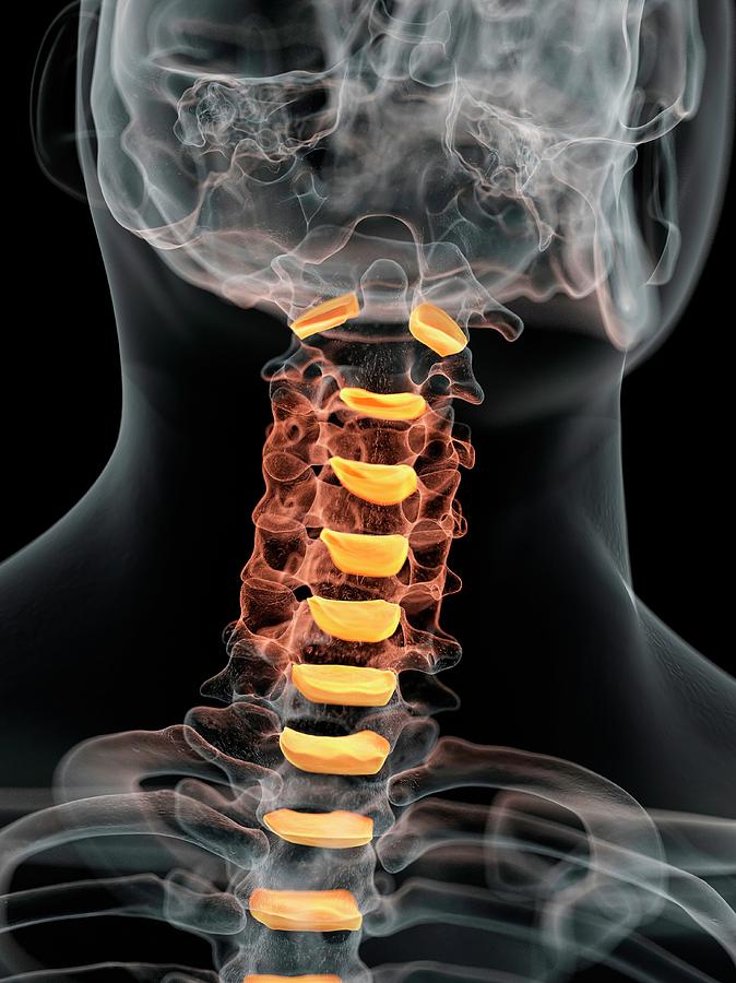
Human Intervertebral Discs Of Neck Photograph by Sciepro Pixels
Definition/Description. The intervertebral disc (IVD) is important in the normal functioning of the spine. It is a cushion of fibrocartilage and the principal joint between two vertebrae in the spinal column. There are 23 discs in the human spine: 6 in the cervical region (neck), 12 in the thoracic region (middle back), and 5 in the lumbar.