Biology Intervertebral disc
Understanding Spinal Anatomy: Intervertebral Discs. Between each vertebral body is a cushion called an intervertebral disc. Each disc absorbs the stress and shock the body incurs during movement and prevents the vertebrae from grinding against one another. The intervertebral discs are the largest structures in the body without a vascular supply.
Classification of Joints · Anatomy and Physiology
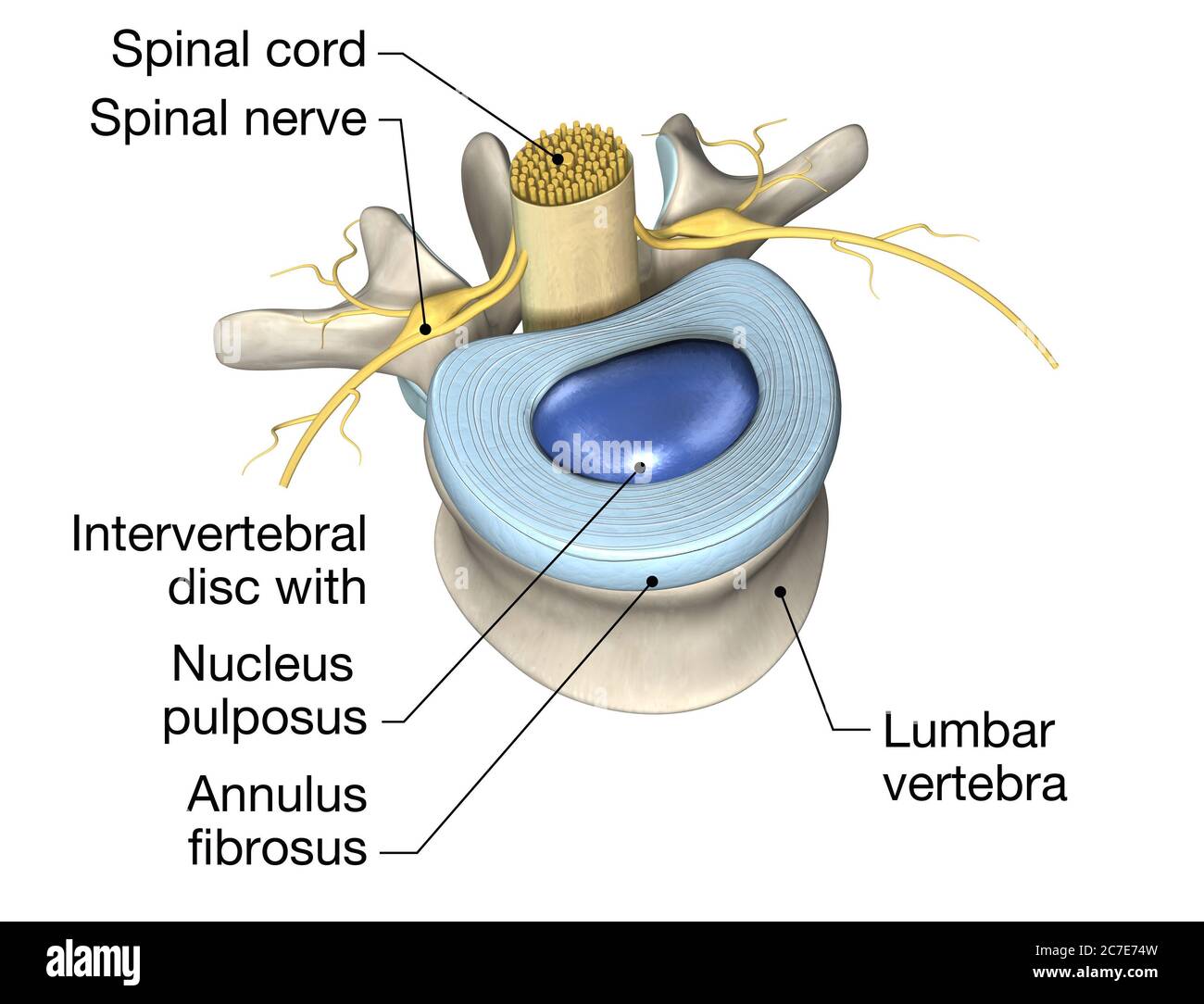
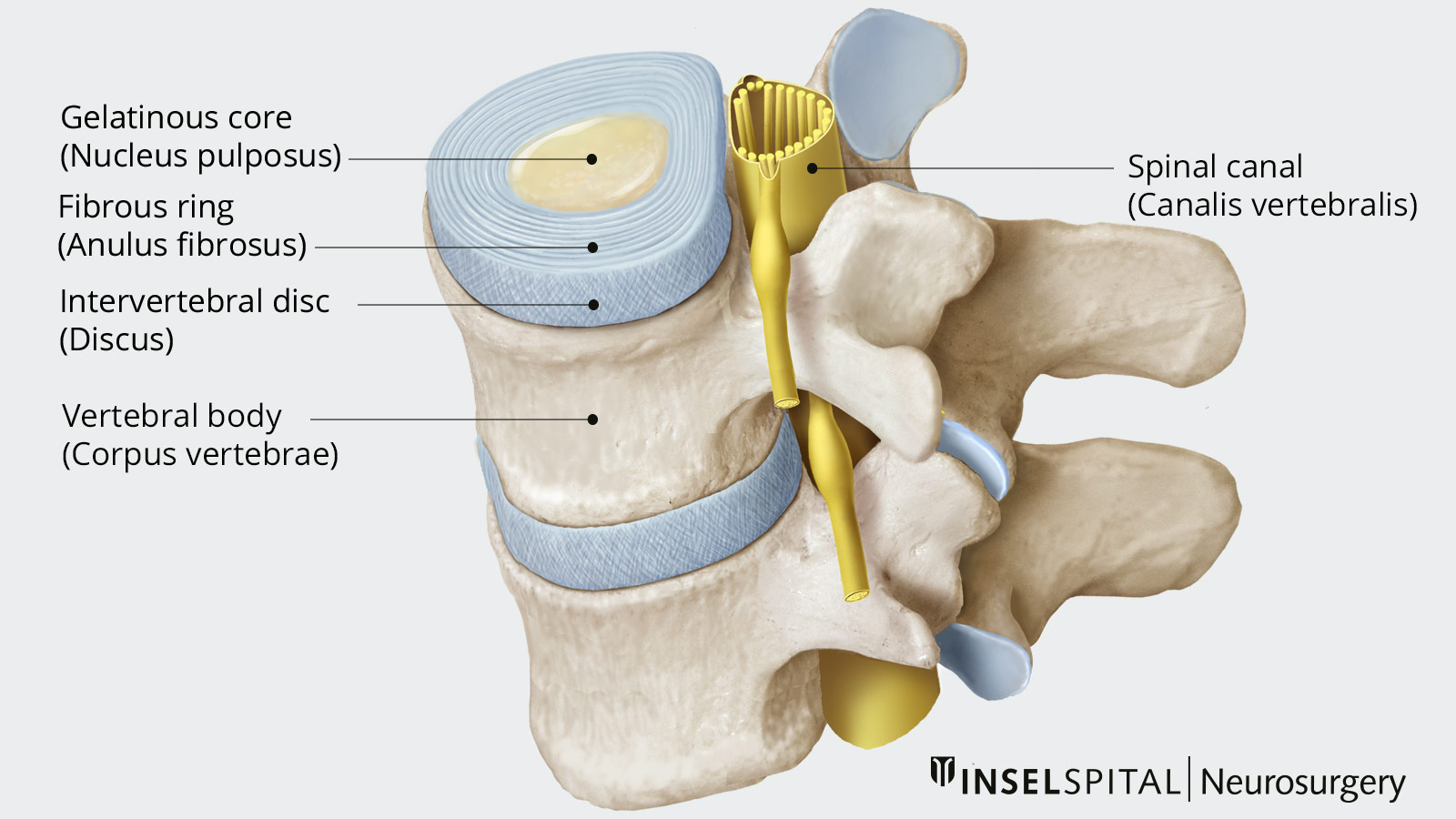
The intervertebral discs are secondary cartilaginous joints, also known as symphyses 7 . Each intervertebral disc is comprised of: peripheral annulus fibrosus. central nucleus pulposus. hyaline cartilage (vertebral side) and fibrocartilage (nucleus pulposus side) Above and below the intervertebral disc are the vertebral body endplates .

Discus intervertebralis with tears and fissures in the annulus... Download Scientific Diagram
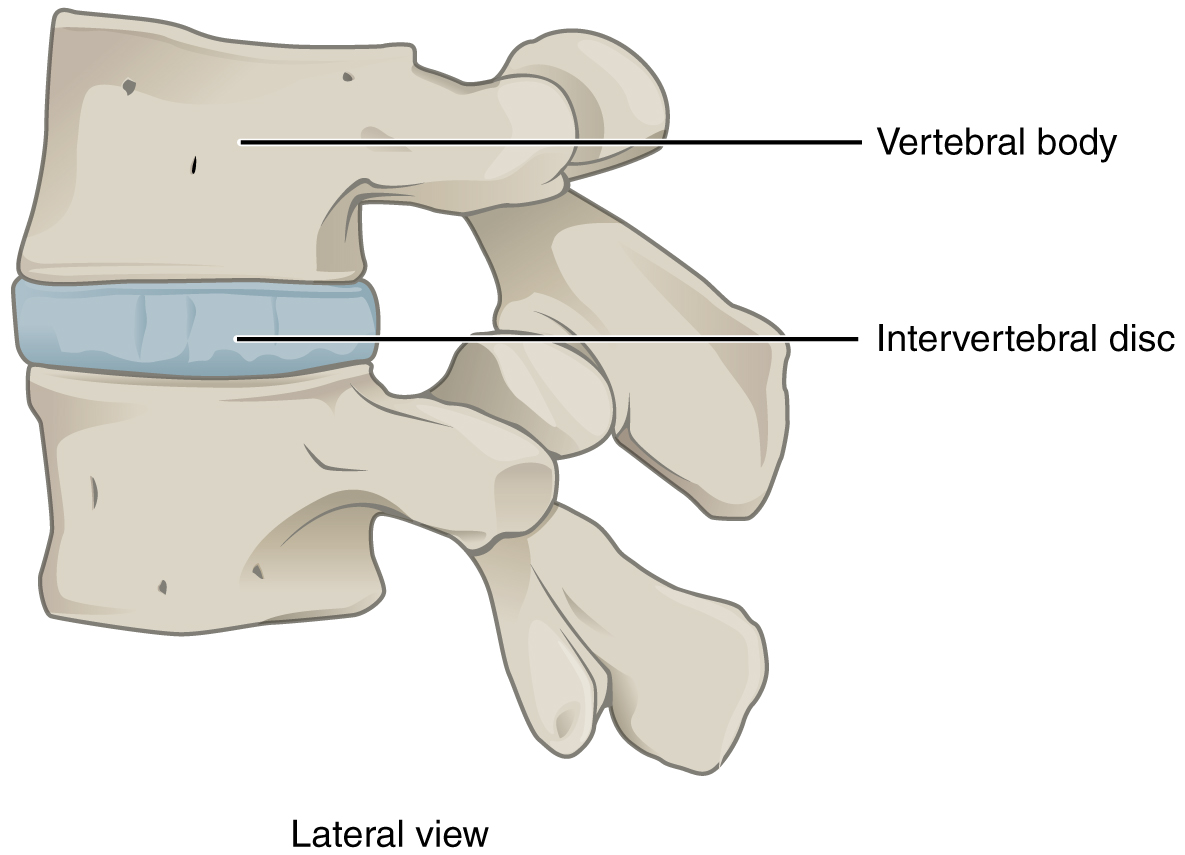
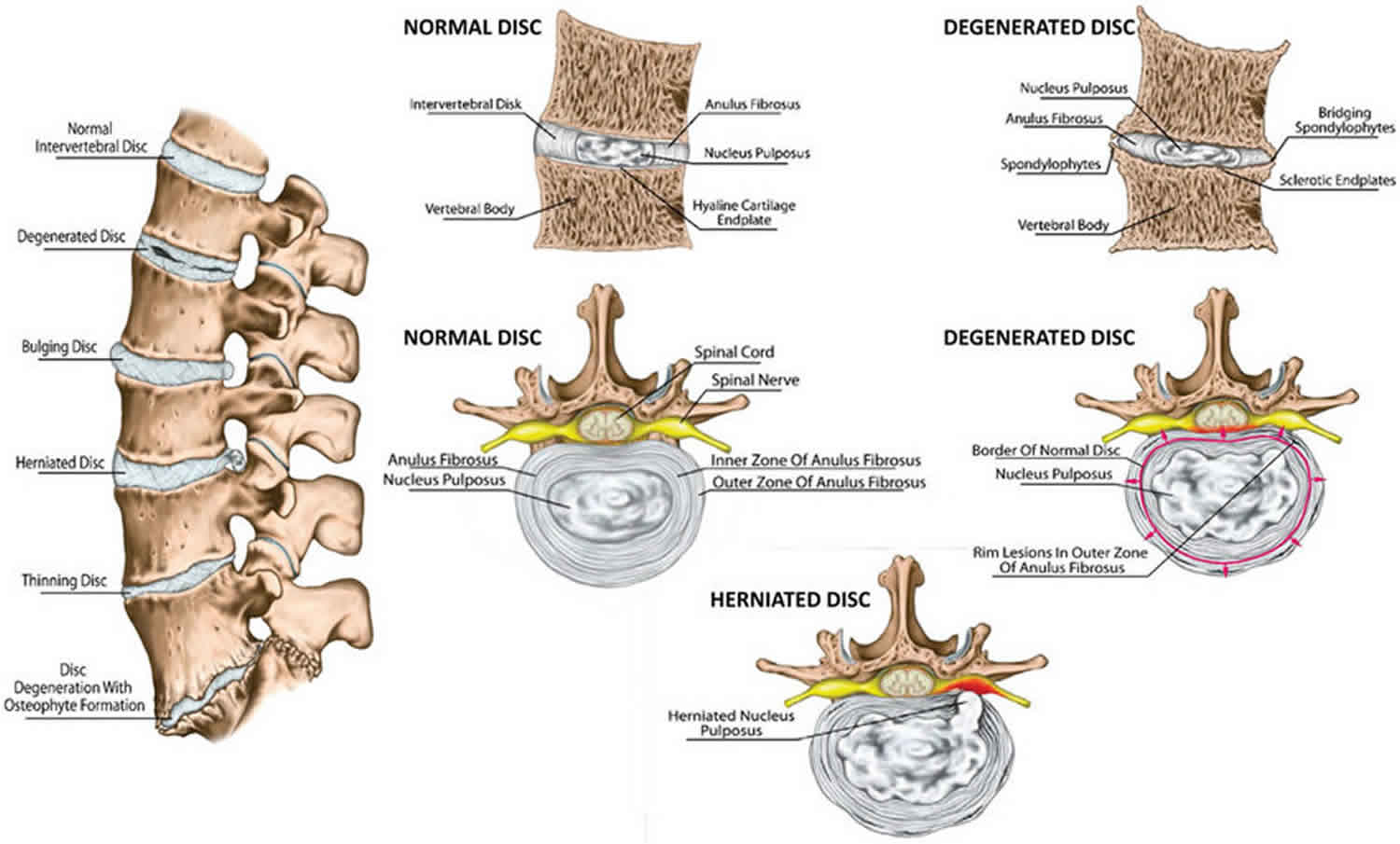
An intervertebral disc is a structure located between adjacent vertebrae of the spine. It consists of a tough outer layer called the annulus fibrosus and a gel-like center called the nucleus pulposus. The intervertebral disc acts as a cushion, allowing the spine to bend and twist without damaging the vertebrae.
Discus intervertebralis T12L1 (Bandscheiben des 12. Brust bis 1. Lendenwirbels) Kenhub
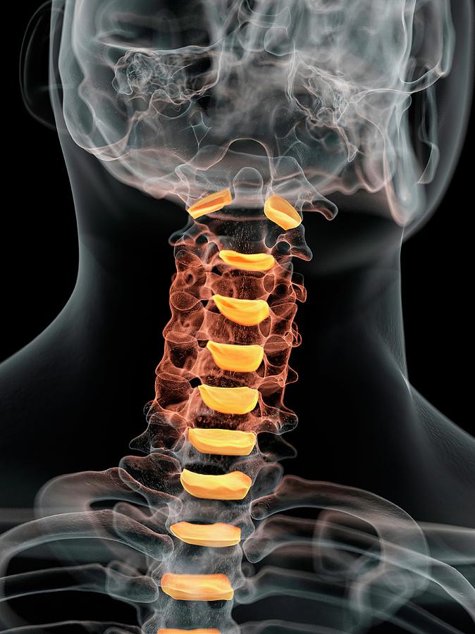
Definition/Description. The intervertebral disc (IVD) is important in the normal functioning of the spine. It is a cushion of fibrocartilage and the principal joint between two vertebrae in the spinal column. There are 23 discs in the human spine: 6 in the cervical region (neck), 12 in the thoracic region (middle back), and 5 in the lumbar.
3D illustration showing lumbal vertebra with intervertebral disc, medically 3D illustration
The intervertebral discs (intervertebral fibrocartilages) are interposed between the adjacent surfaces of the bodies of the vertebræ, from the axis to the caudal vertebrae, and form the chief bonds of connection between the vertebræ.They vary in shape, size, and thickness, in different parts of the vertebral column, the thickness decreasing through the thoracic and lumbar region.Each.
Human Intervertebral Discs Of Neck Photograph by Sciepro Pixels
Ein Discus intervertebralis kann in zwei Teile unterteilt werden, den äußeren Faserring (Anulus fibrosus) und den innen liegenden Gallertkern (Nucleus pulposus).Anulus fibrosus. Der äußere, derbe Teil des Discus intervertebralis besteht aus Faserknorpel.Die Lamellen dieses straffen kollagenen Bindegewebes sind konzentrisch geschichtet und strahlen in Deckplatte und Randleiste der.

Diskus intervertebralis II Anatomi og Fysiologi
Discus intervertebralis. Definition. The intervertebral discs (intervertebral fibrocartilages) are interposed between the adjacent surfaces of the bodies of the vertebræ, from the axis to the sacrum, and form the chief bonds of connection between the vertebræ. They vary in shape, size, and thickness, in different parts of the vertebral column

Anatomy Stock Images neckintervertebraldiscdiscusintervertebralisspinousprocessfacet
The structural elements, both macroscopically and microscopically, together with the biochemical elements, are intimately related to function. The intervertebral disc should not be though of as a homogeneous and static structure; it has a heterogeneous composition and responds dynamically to applied loads. Neither should it be considered as an.

Anatomy Stock Images neckintervertebraldiscdiscusintervertebralisspinousprocessfacet
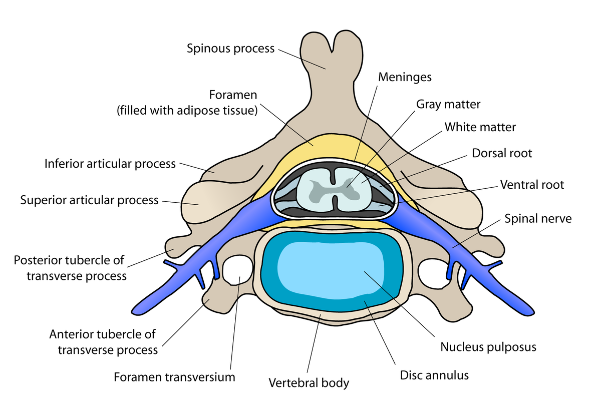
Intervertebral discs consist of an outer fibrous ring, the anulus (or annulus) fibrosus disci intervertebralis, which surrounds an inner gel-like center, the nucleus pulposus. The anulus fibrosus consists of several layers (laminae) of fibrocartilage made up of both type I and type II collagen.Type I is concentrated toward the edge of the ring, where it provides greater strength.
Intervertebral Disc Anatomy
Discus intervertebralis 1/3. Synonyms: Intervertebral fibrocartilage, Fibrocartilago intervertebralis The articular surfaces of directly adjacent vertebral bodies are separated by fibrocartilaginous intervertebral discs. They adhere to both the vertebral end-plates and the bony vertebral rim. The attachment to the vertebral bodies is via ring.

Anatomy Stock Images neckintervertebraldiscdiscusintervertebralisspinousprocess
This review article describes anatomy, physiology, pathophysiology and treatment of intervertebral disc. The intervertebral discs lie between the vertebral bodies, linking them together. The components of the disc are nucleus pulposus, annulus fibrosus and cartilagenous end-plates. The blood supply to the disc is only to the cartilagenous end.
Lumbar vertebra with intervertebral disc, medically 3D illustration ilustração do Stock Adobe
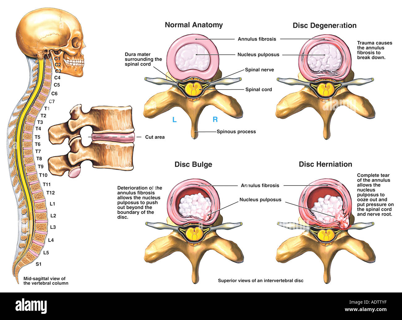
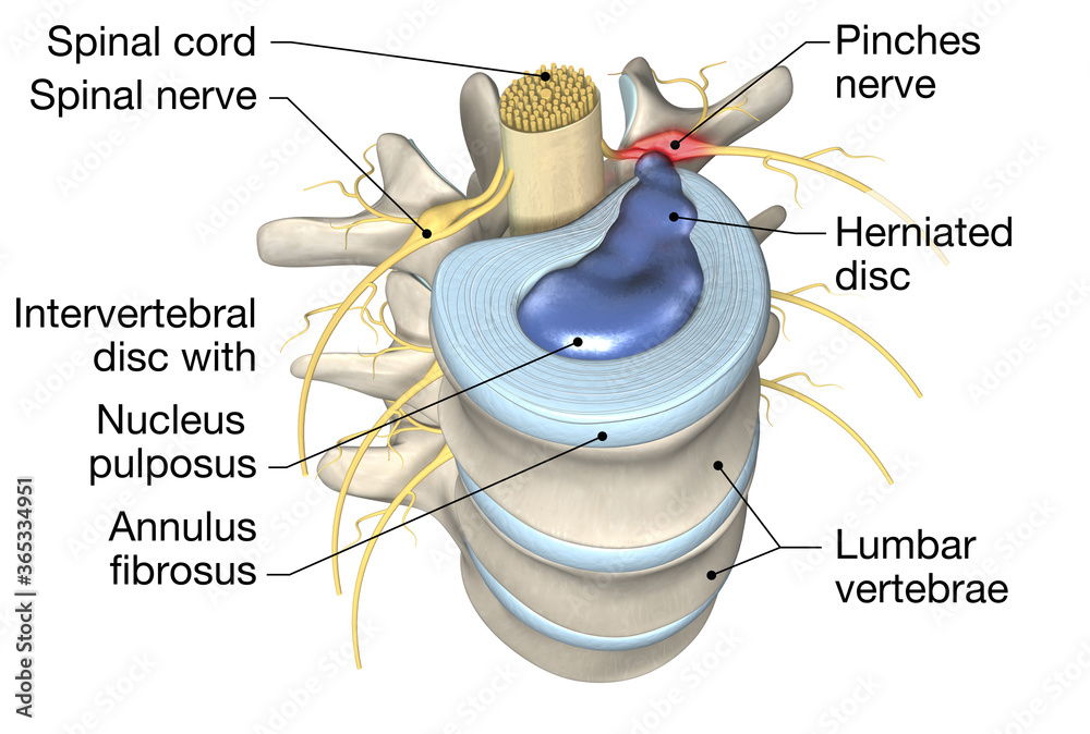
Intervertebral disc disease is a common condition characterized by the breakdown (degeneration) of one or more of the discs that separate the bones of the spine (vertebrae), causing pain in the back or neck and frequently in the legs and arms. The intervertebral discs provide cushioning between vertebrae and absorb pressure put on the spine.
Intervertebral discs Anatomy and embryology Kenhub
This review begins with a brief introduction in which the development, blood supply and innervation of the intervertebral disc is considered, particularly as these may influence the following sections on structure and function.
Herniated Disc Neurosurgery Inselspital Bern
Discus intervertebralis 1/4. Synonyms: Intervertebral fibrocartilage, Fibrocartilago intervertebralis After learning about individual vertebrae, it's time to explore how the vertebral column is kept together as a unit. Adjacent vertebral bodies are joined by symphyses called intervertebral symphyses (discs). The exceptions are C1-C2 and after.
Intervertebral disc Wikipedia
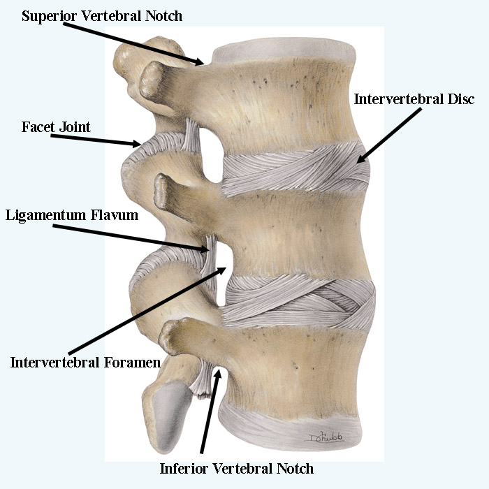
Adjacent vertebrae articulate through zygapophyseal joints between the respective superior and inferior facets of the vertebral articular processes as well as through the joints of the vertebral bodies. While the former serves to limit the spine's range of motion, the latter increases it and provides the majority of the spine's weight-bearing capacity. The inferior surface of the superior.
Intervertebral disc anatomy, function, degeneration, herniation
Introduction. Back pain is a major public health problem in Western industrialized societies. It causes suffering and distress to patients and their families, and affects a large number of people; the point prevalence rates in a number of studies ranged from 12% to 35% [], with around 10% of sufferers becoming chronically disabled.It also places an enormous economic burden on society; its.