Foot Pain Diagram exatin.info
Find the Components You Need. Free UK Delivery on Eligible Orders!
Lisfranc Injuries Core EM
The foot is a very complex part of the body. It is made up of a great many bones, joints, muscles, nerves, and tendons. The foot has two important jobs to perform for the body. Those jobs are to propel the body forward and to absorb shock. Fortunately, the foot is perfectly designed to do those jobs.

anatomy of the foot Ballet News Straight from the stage bringing
33 joints more than 100 muscles, tendons, and ligaments Bones of the foot The bones in the foot make up nearly 25% of the total bones in the body, and they help the foot withstand weight..

Labeled Diagram Of The Foot Teenage Lesbians
Foot Anatomy There are many parts of the foot and all have important jobs. Each foot has 26 bones, over 30 joints, and more than 100 muscles, ligaments, and tendons. These structures work together to carry out two main functions: Bearing weight Forward movement (propulsion)
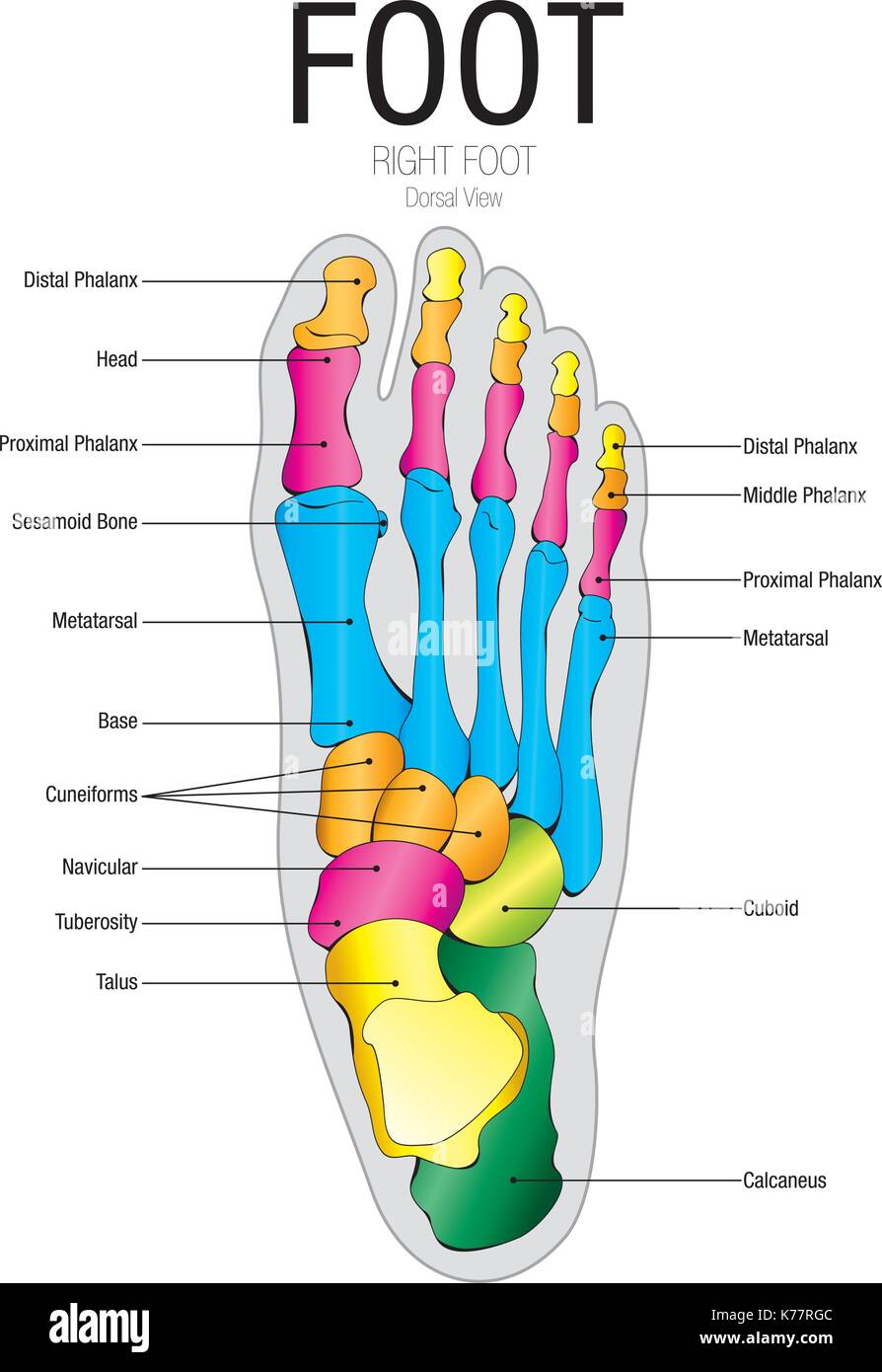
Chart of FOOT Dorsal view with parts name Vector image Stock Vector
The foot is the region of the body distal to the leg that is involved in weight bearing and locomotion. It consists of 28 bones, which can be divided functionally into three groups, referred to as the tarsus, metatarsus and phalanges. The foot is not only complicated in terms of the number and structure of bones, but also in terms of its joints.
Notes on Anatomy and Physiology Using Imagery to Relax the Weight
There are a variety of anatomical structures that make up the anatomy of the foot and ankle (Figure 1) including bones, joints, ligaments, muscles, tendons, and nerves. These will be reviewed in the sections of this chapter. Figure 1: Bones of the Foot and Ankle Regions of the Foot
Advantage Orthopedic and Sports Medicine Clinic Gresham, OR Health
These arches — the medial arch, lateral arch, and fundamental longitudinal arch — are created by the angles of the bones and strengthened by the tendons that connect the muscles and the ligaments.

The anatomy of the foot Schuhdealer blogA blog all about sneakers and
It is made up of three joints: upper ankle joint (tibiotarsal), talocalcaneonavicular, and subtalar joints. The last two together are called the lower ankle joint. The upper ankle joint is formed by the inferior surfaces of tibia and fibula, and the superior surface of talus.
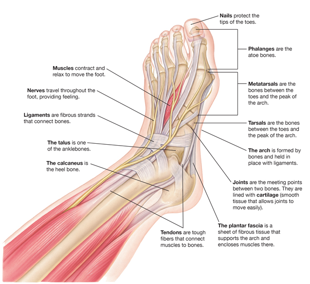
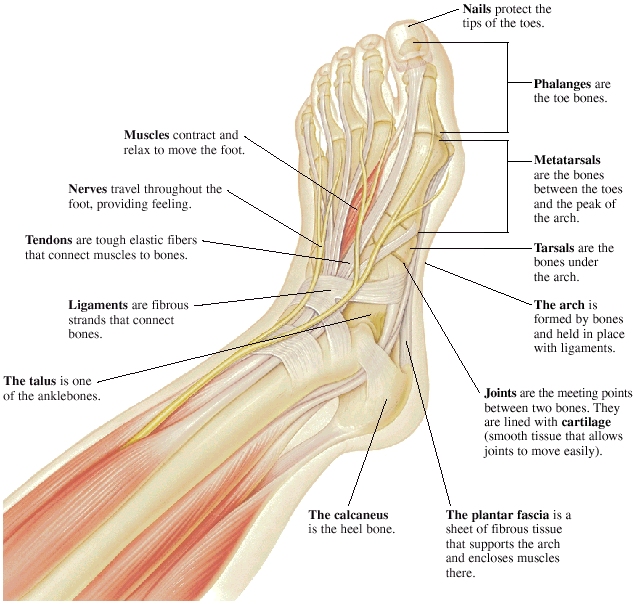
Parts of a Foot Saint Luke's Health System
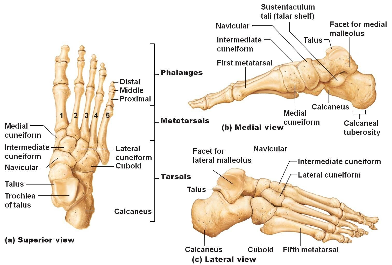
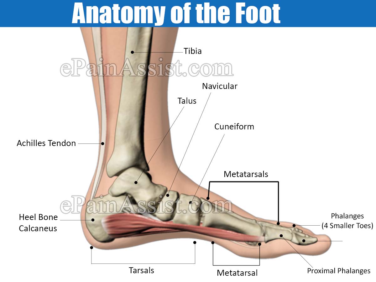
Bones of foot. The 26 bones of the foot consist of eight distinct types, including the tarsals, metatarsals, phalanges, cuneiforms, talus, navicular, and cuboid bones. The skeletal structure of.
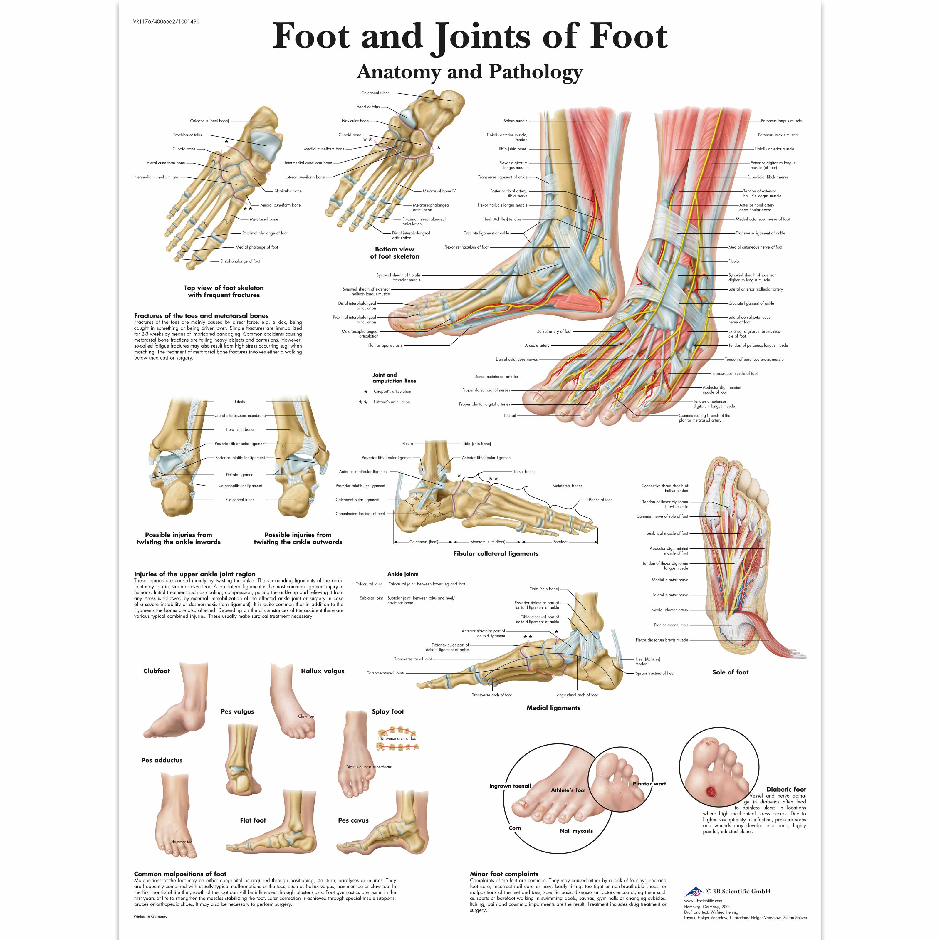
Anatomical Charts and Posters Anatomy Charts Foot and Ankle
The Toes, Arch and Heel Toes are the parts of the foot that allow people to move. They help people grip the ground and push off when they walk or run. The arch is the part of the foot that helps to absorb shock when we move around. It is located between the heel and the toes. The heel provides balance and stability.
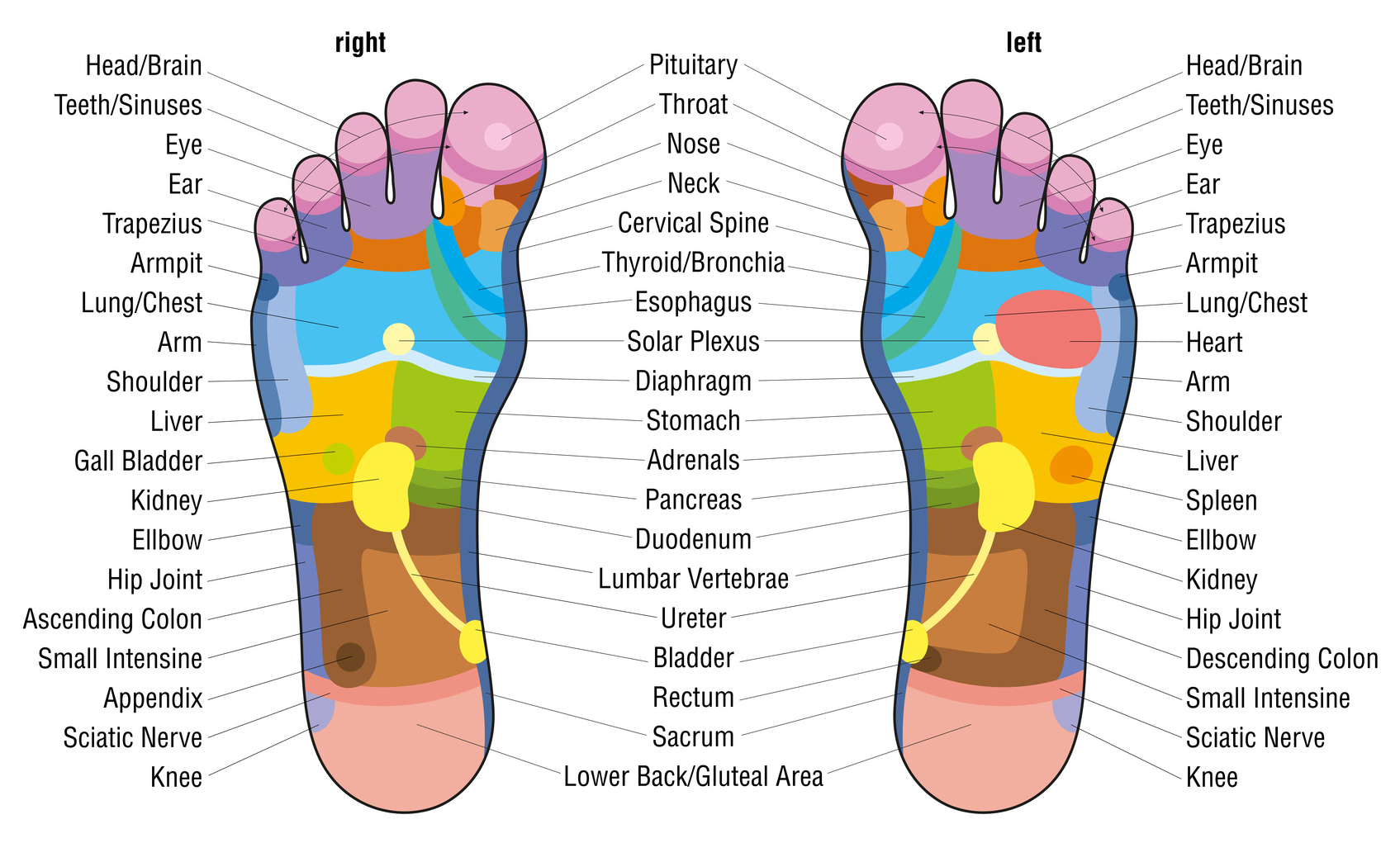
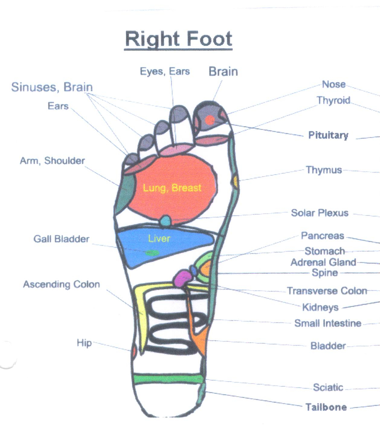
What is foot massage? Urban Blog
Foot Anatomy The foot contains 26 bones, 33 joints, and over 100 tendons, muscles, and ligaments. This may sound like overkill for a flat structure that supports your weight, but you may not realize how much work your foot does!

Anatomy of human foot with labels on white background — ankle, leg
These bones are arranged in two rows, proximal and distal. The bones in the proximal row form the hindfoot, while those in the distal row from the midfoot. Hindfoot. Talus. Calcaneus. The talus connects the foot to the rest of the leg and body through articulations with the tibia and fibula, the two long bones in the lower leg. Midfoot. Navicular.
Foot Pain & Its Anatomical DistributionCauses of Foot Pain
Joints Big toe Ball of foot Arch Heel General pain and swelling Speaking with a doctor Summary The location of pain in the foot can sometimes indicate the underlying cause. The cause will.

Human Foot Bones Labeled
Ligaments are fibrous strands that connect bones. Nerves travel throughout the foot, providing feeling. Nails protect the tips of the toes. Phalanges are the toe bones. Metatarsals are the bones between the toes and the ankle bones. Tarsals are bones of the rear foot (hindfoot) or middle foot (midfoot). The talus is one of the ankle bones.

This chart shows foot and ankle bone and ligament anatomy, normal
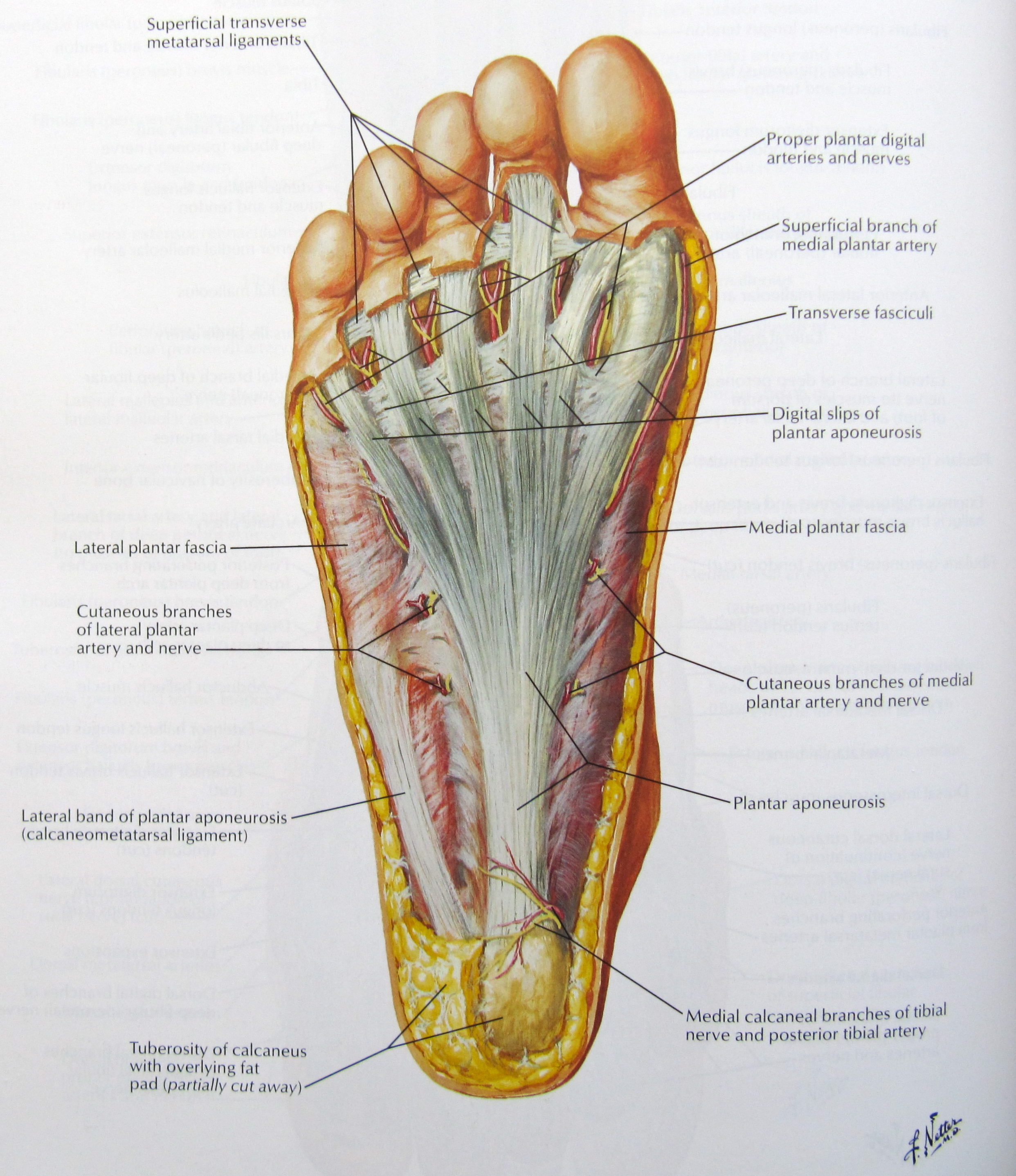
The foot can be divided into two main parts - the sole or plantar region, which is the part of the foot contacting the ground, and the dorsum of the foot or the dorsal region, which is the part directed superiorly. Alternatively, it can be divided into three sections.
Foot Pain Natural Relief and Herbal Remedies HubPages
Human feet allow bipedal locomotion, and they are an essential sensory structure for postural control. The foot structure is complex, consisting of many bones, joints, ligaments and muscles. The foot is divided into three parts: rearfoot, midfoot, and forefoot. A clinician's ability to understand the anatomical structures of the foot is crucial.