
BrainMidsagittal Section Diagram Quizlet
Twenty-Five Diagnoses on Midline Images of the Brain: From Fetus to Child to Adult. 2018 Jan-Feb;38 (1):218-235. doi: 10.1148/rg.2018170019. Midsagittal images of the brain provide a wealth of anatomic information and may show abnormalities that are pathognomonic for particular diagnoses. Using an anatomy-based approach, the authors identify.

Midsagittal Section Of The Brain bmpwabbit
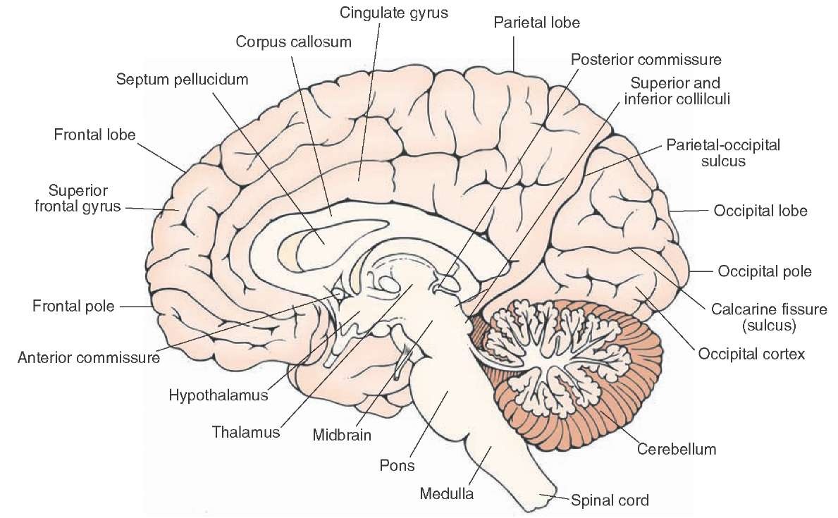
When the brain is hemisected in the midsagittal plane, all of its major subdivisions plus a number of additional structures are visible on the cut surface (Figure 1.14). In this view, the cerebral hemispheres, because of their great size, are still the most obvious structures. The frontal lobe of each hemisphere extends forward from the central sulcus, the medial end of which can just be seen.

Midsagittal Section Of The Human Brain Anatomy Body List Human Brain
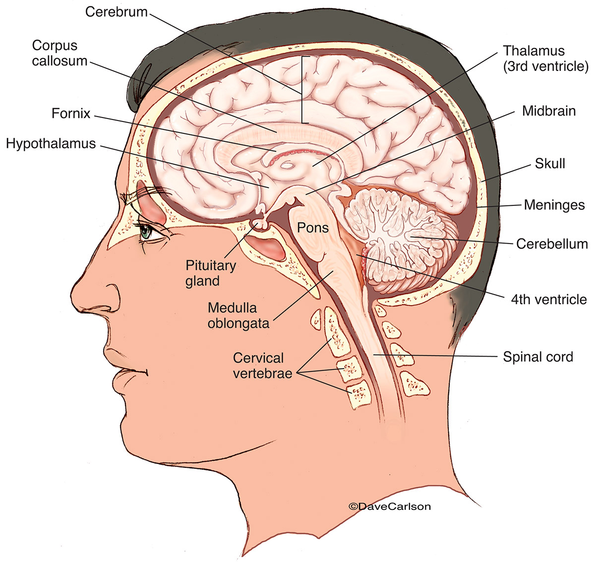
The thalamus is located deep in the brain. It can be seen in the midsagittal section in the area of the third ventricle. Many pathways between the cerebrum, brainstem, cerebellum, and spinal cord have a synapse in the thalamus.. The UBC Functional Anatomy images show the white and grey matter with its typical coloration. A list of the.
Human Brain Midsagittal View Carlson Stock Art
Previous analyses have been published which investigate the brain midsagittal shape variation in adult humans using digital anatomy and geometric morphometrics, this plane being relevant in terms of biological organization and human evolution (Bruner et al. 2010, 2014a). However, we will ignore how these brain morphological variations can.
Midsagittal Brain Diagram My XXX Hot Girl
All components of the ventricular system, except perhaps for the lateral ventricles, can be seen on a typical medial surface of the brain cut in the midsagittal plane. In Figure 1.12 , the lateral ventricle is visible in this hemisphere because the septum pellucidum has been dissected away; this is a very thin structure made of ependymal cells.

Midsagittal Section Of The Human Brain Chapter 12 The Central
brainstem are visible on the medial surface of a brain that has been cut in the midsagittal plane. Parts of all of the subdivisions are also visible from the ventral surface of the whole brain. In this set of tutorials, you will find video demonstrations (from the brain anatomy lab) and photographs (in the tutorial notes)

Midsagittal Section Of The Brain Diagram Ajor Png
Midsagittal images of the brain provide a wealth of anatomic information and may show abnormalities that are pathognomonic for particular diagnoses. Using an anatomy-based approach, the authors identify pertinent anatomic structures to serve as a checklist when evaluating these structures. Subregions evaluated include the corpus callosum, pituitary gland and sellar region, pineal gland and.
Overview of the Central Nervous System (Gross Anatomy of the Brain) Part 1
This preview gives a sneak peek to our tutorial on the anatomy seen on a medial view of the brain, also known as a midsagittal section of the brain.Take a cl.

Midsagittal Section Of The Brain bmpwabbit
Brain, midsagittal view. Three views of brainstem. Top and anterior views of cerebellum. Major nuclei of thalamus. Lateral and medial surfaces of cerebrum, showing major sulci and gyri.. Anatomy Brain Anatomy; 2003/viewarticle/998119. A New Era in the Management of GERD 1.0 CME / CE / ABIM MOC Credits.

brain midsagittal view labels
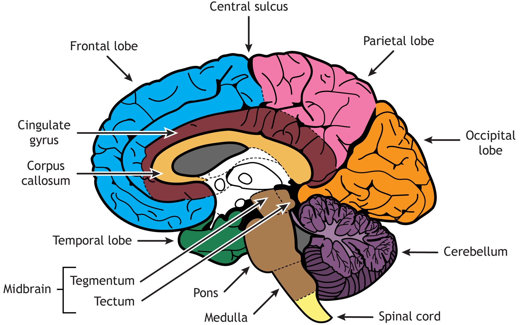
The midsagittal section of the brain shows the three major parts of the brain, which are the cerebrum, cerebellum, and brainstem. The cerebrum (prosencephalo.
Midsagittal section of the brain Anatomy Kenhub
In this study, midsagittal brain shape variation is investigated in a sample of 102 humans, in order to describe and quantify the major patterns of correlation between morphological features, the effect of size and sex on general anatomy, and the degree of integration between different cortical and subcortical areas.
Brain Structure Differentiation Introduction to Neuroscience
the sagittal midline is observed when the cerebral aqueduct can be seen draining the third ventricle into the fourth ventricle. any displacement, of the cerebellar tonsils, or crowding of the foramen magnum. a review of the cisterns is important to note any displacement of the midline. moving superiorly, the cerebral aqueduct is observed for.

sagittal view of the human brain BRAIN SAGITTAL Anatomy
Welcome to the Midsagittal Brain Study Module Page! Below you will find links to modules designed to help you learn all about the structures of the brain visible from a midsagittal section. In the Human Brain Anatomy Study Module , the parts of the brain are taken apart and put back together to help teach you about the structure and function of.
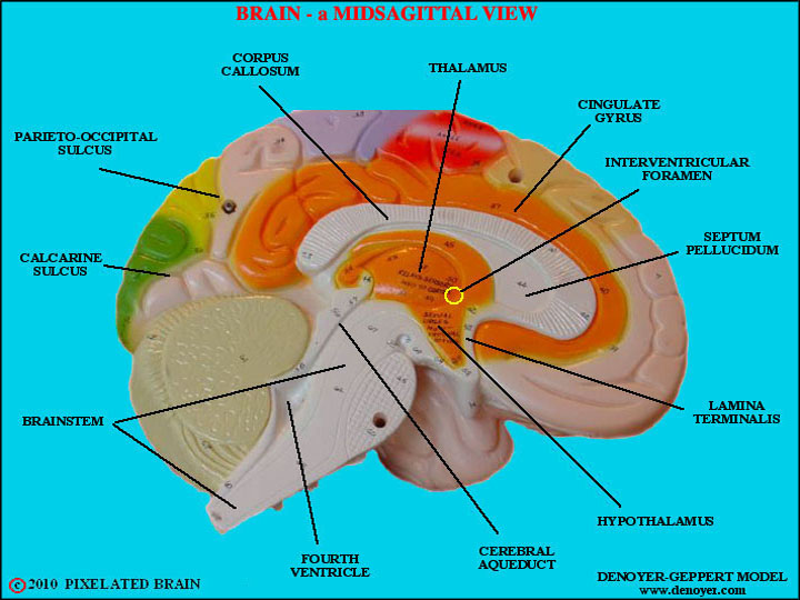
Pixelated Brain a model showing a midsagittal view of the brain
Figure 18.1. A midsagittal section of the brain. All four cerebral lobes are visible, as in the cingulate gyrus, which extends through the medial aspects of the frontal and parietal lobes. The corpus callosum sits beneath the cingulate gyrus. Below the cerebrum lies the midbrain, pons, medulla and cerebellum.

Midsagittal view of brain with labelled structures … Brain Anatomy
3. Mid-sagittal aspect of the brain. Now, let's briefly turn our attention to a single cerebral hemisphere. When the brain is cut in the midsagittal plane, all of its subdivisions are visible on the cut surface (see Figure 1.5B). Just as in the embryo, the subdivisions are arranged as though they were stacked, with the cerebral hemisphere.

Midsagittal section of the brain. The Central Nervous System
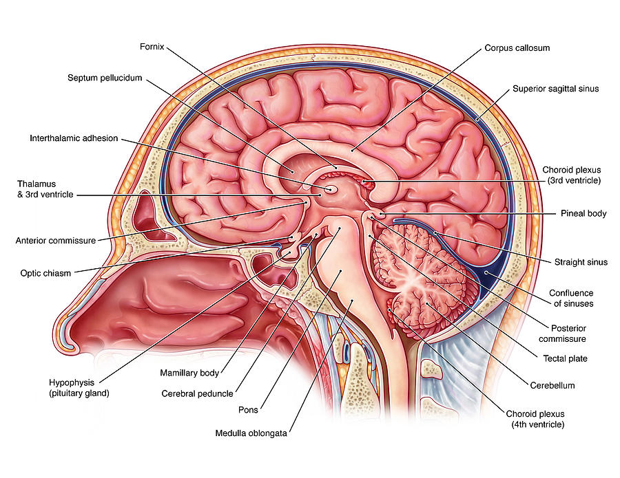
Midsagittal section of the deep brain anatomy. This midline view demonstrates the third ventricle, with its roof formed by the body and column of the fornices and the velum interpositum. In the midline anteriorly, the lamina terminalis, optic chiasm and pituitary infundibulum are visible. The floor of the third ventricle is composed of the.