Muscles that lift the Arches of the Feet
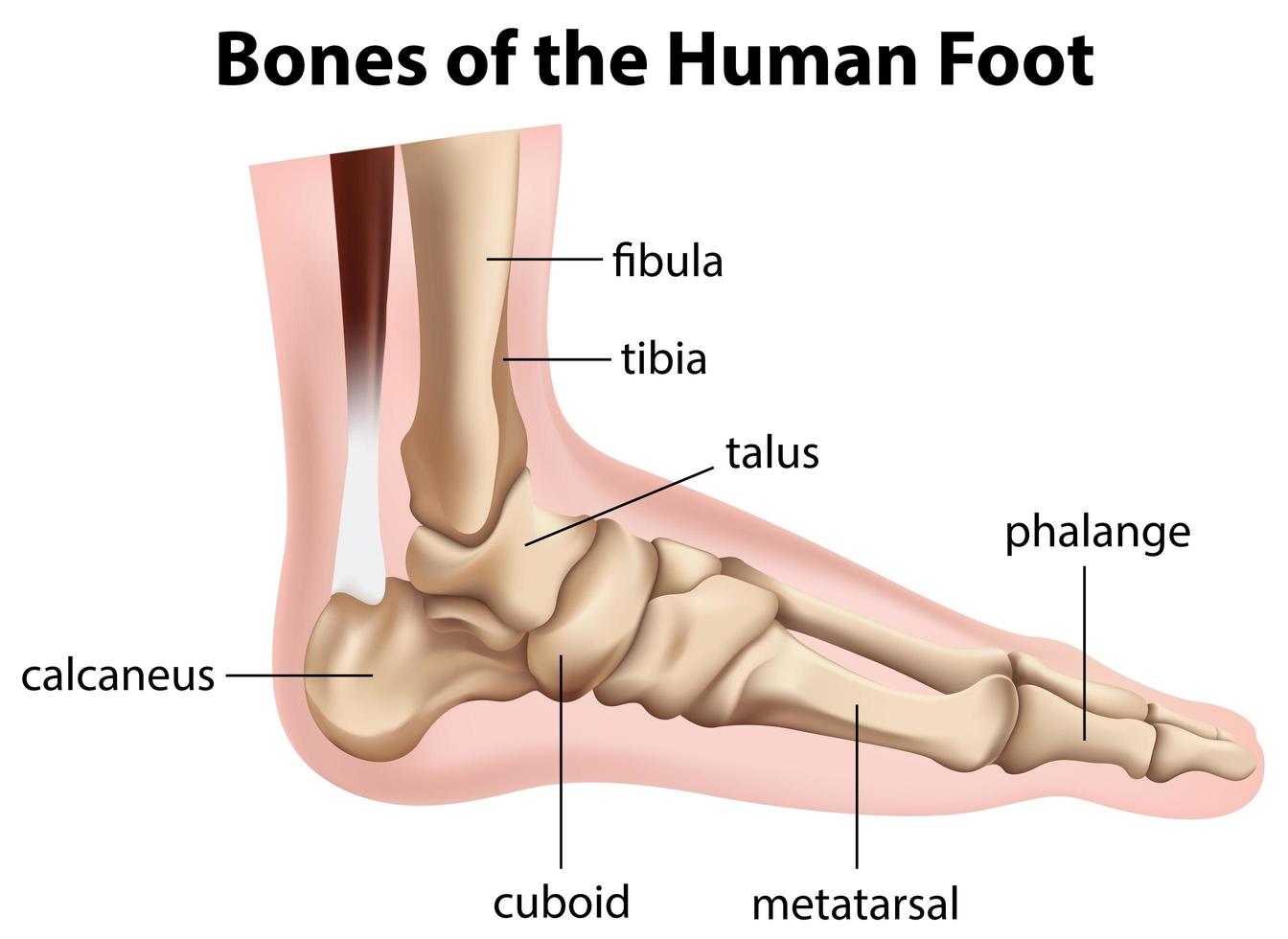
The foot can also be divided up into three regions: (i) Hindfoot - talus and calcaneus; (ii) Midfoot - navicular, cuboid, and cuneiforms; and (iii) Forefoot - metatarsals and phalanges. In this article, we shall look at the anatomy of the bones of the foot - their bony landmarks, articulations, and clinical correlations.
Bones of the human foot diagram 1142236 Vector Art at Vecteezy
The anatomy of the foot The foot contains a lot of moving parts - 26 bones, 33 joints and over 100 ligaments. The foot is divided into three sections - the forefoot, the midfoot and the hindfoot. The forefoot This consists of five long bones (metatarsal bones) and five shorter bones that form the base of the toes (phalanges).
Human Anatomy for the Artist The Dorsal Foot How Do I Love Thee? Let
Figure 1: Bones of the Foot and Ankle Regions of the Foot The foot is traditionally divided into three regions: the hindfoot, the midfoot, and the forefoot (Figure 2). Additionally, the lower leg often refers to the area between the knee and the ankle and this area is critical to the functioning of the foot.

Anatomy of the Foot and Ankle OrthoPaedia
Maverick biologist explains the human condition and makes sense of life. Watch brilliant biologist solve the human condition & finally explain the meaning of life.

Foot and Ankle Musculoskeletal Key
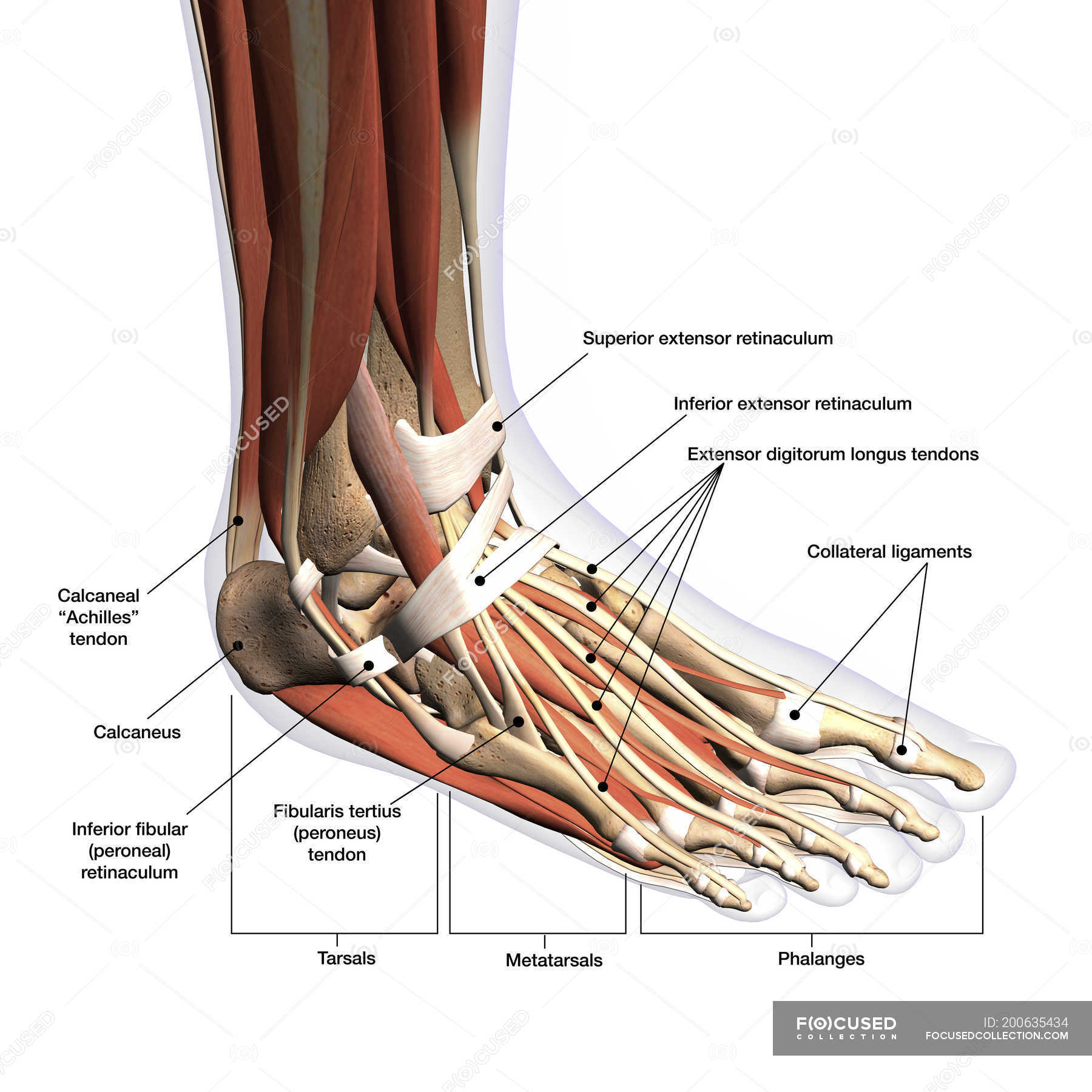
The foot is the region of the body distal to the leg and consists of 28 bones. These bones are arranged into longitudinal and transverse arches with the support of various muscles and ligaments. There are three arches in the foot, which are referred to as: Medial longitudinal arch Lateral longitudinal arch Transverse arch

Pin on Medical Podiatry
Morton's neuroma is a common foot problem where compression on a nerve in the ball of the foot causes burning, tingling, and pain near the third and fourth toes. It can make you feel like you have a pebble in your shoe or on a fold in your sock. Wearing high heels is a common cause of Morton's neuroma.

image lateral_ankle for term side of card Ligament Tear, Ligaments And
The talus is held in place by the foot bones surrounding it and various ligaments. 4. Calcaneus. The calcaneus is more commonly known as the heel bone. It is the largest of the foot bones and has a quadrangular shape. The calcaneus is the most commonly fractured tarsal bone, usually from a high fall.
.jpg)
Foot Bone Diagram resource Imageshare
A foot pain diagram is a great tool to help you work out what is causing your ankle and foot pain. There are a whole range of structures e.g. bones, muscles, tendons and nerves which will each give slightly different foot pain symptoms.
Turf toe causes, signs, symptoms, recovery, diagnosis & turf toe treatment
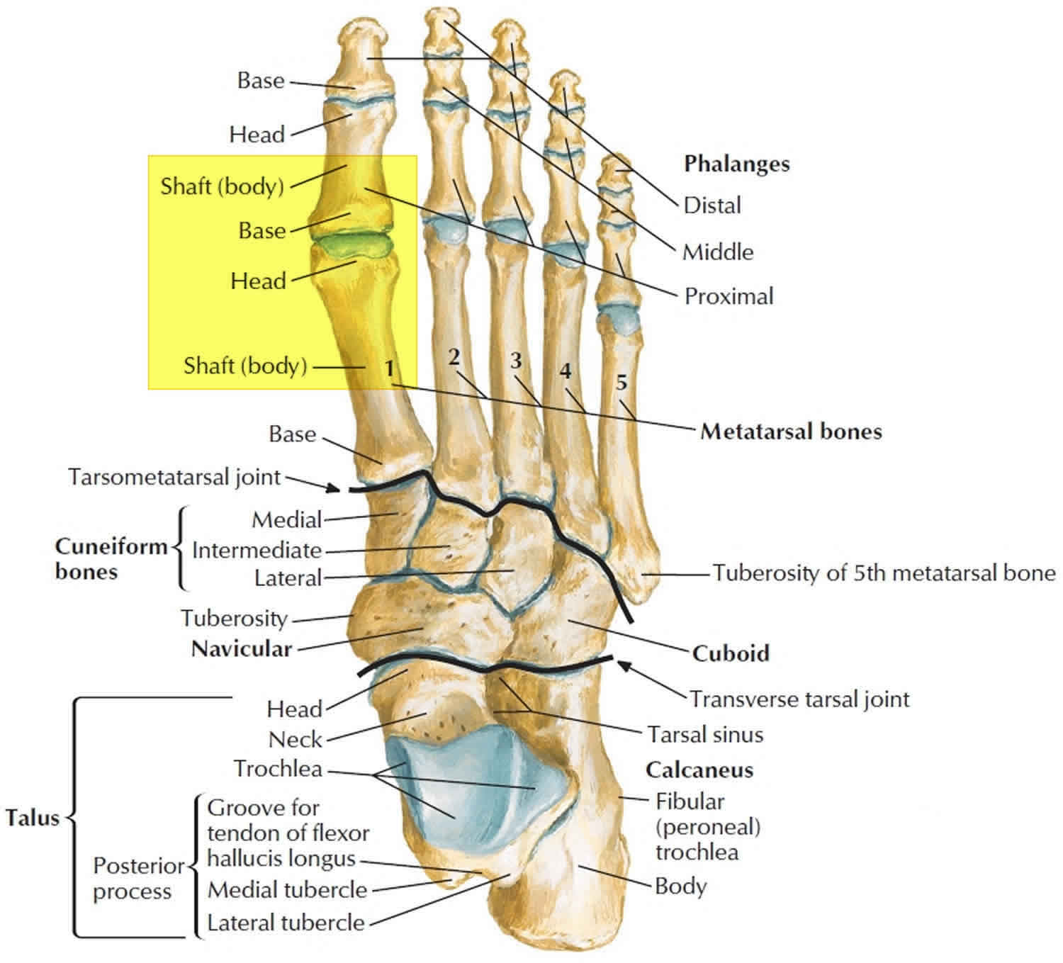
Phalanges Also known as toe bones, these are the 14 long bones in the toes on each foot. As mentioned above, these form the forefoot with the metatarsals. The second to fifth toes have 3 phalanges each, while only 2 are located in the big toe.

Ankle and Foot Pain Massage Therapy Connections
Foot Bones: Forefoot. The forefoot consists of 19 bones; 5 metatarsal bones and 14 phalanges. The big toe has 2 phalanges bones, while the remaining four have 3 phalanges each. The 1st metatarsal is the shortest and thickest of the metatarsals, and it is designed to take up to 40% of your body weight in standing, which rises to 70% when walking.
.jpg)
Foot Bone Diagram resource Imageshare
33 joints more than 100 muscles, tendons, and ligaments Bones of the foot The bones in the foot make up nearly 25% of the total bones in the body, and they help the foot withstand weight..

Buy Human Foot and Ankle Anatomy Chart Online at Low Prices in India
Foot bones and anatomy Conditions affecting the foot bones When to see a doctor Summary The foot is an intricate part of the body, consisting of 26 bones, 33 joints, 107 ligaments, and 19.
Anatomy of human foot with labels on white background — ankle, leg
Dr. Ebraheim's educational animated video describes anatomical structures of the foot and ankle, The Bony Anatomy, The Joints, Ligaments, and the Compartments, in a simple and easy way..more.

Diagrams of Foot 101 Diagrams
The distal phalanges (foot) are located at the end of each toe. Three phalangeal bones make up each digit, articulating with each other at bending joints. The distal phalanges come at the end.

Foot and Ankle Anatomical Chart Anatomy Models and Anatomical Charts
There are 26 bones in the foot, divided into three groups: Seven tarsal bones Five metatarsal bones Fourteen phalanges Tarsals make up a strong weight bearing platform. They are homologous to the carpals in the wrist and are divided into three groups: proximal, intermediate, and distal.
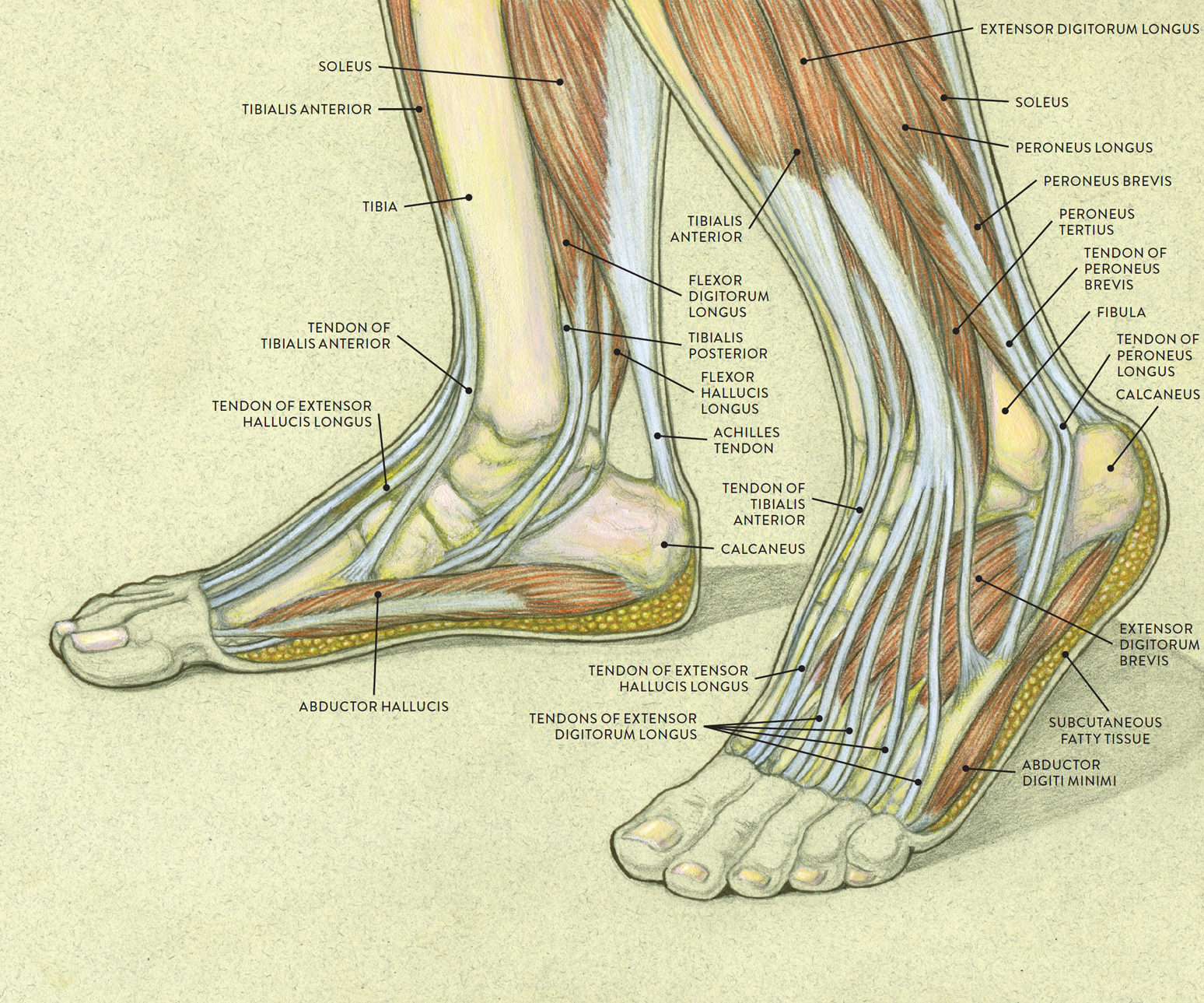
Muscles of the Leg and Foot Classic Human Anatomy in Motion The
Foot Anatomy, Pictures & Model | Body Maps Human body Foot Foot The foot is the lowermost point of the human leg. The foot's shape, along with the body's natural balance-keeping systems,.