Brain sections and organ part functions in labeled anatomical outline
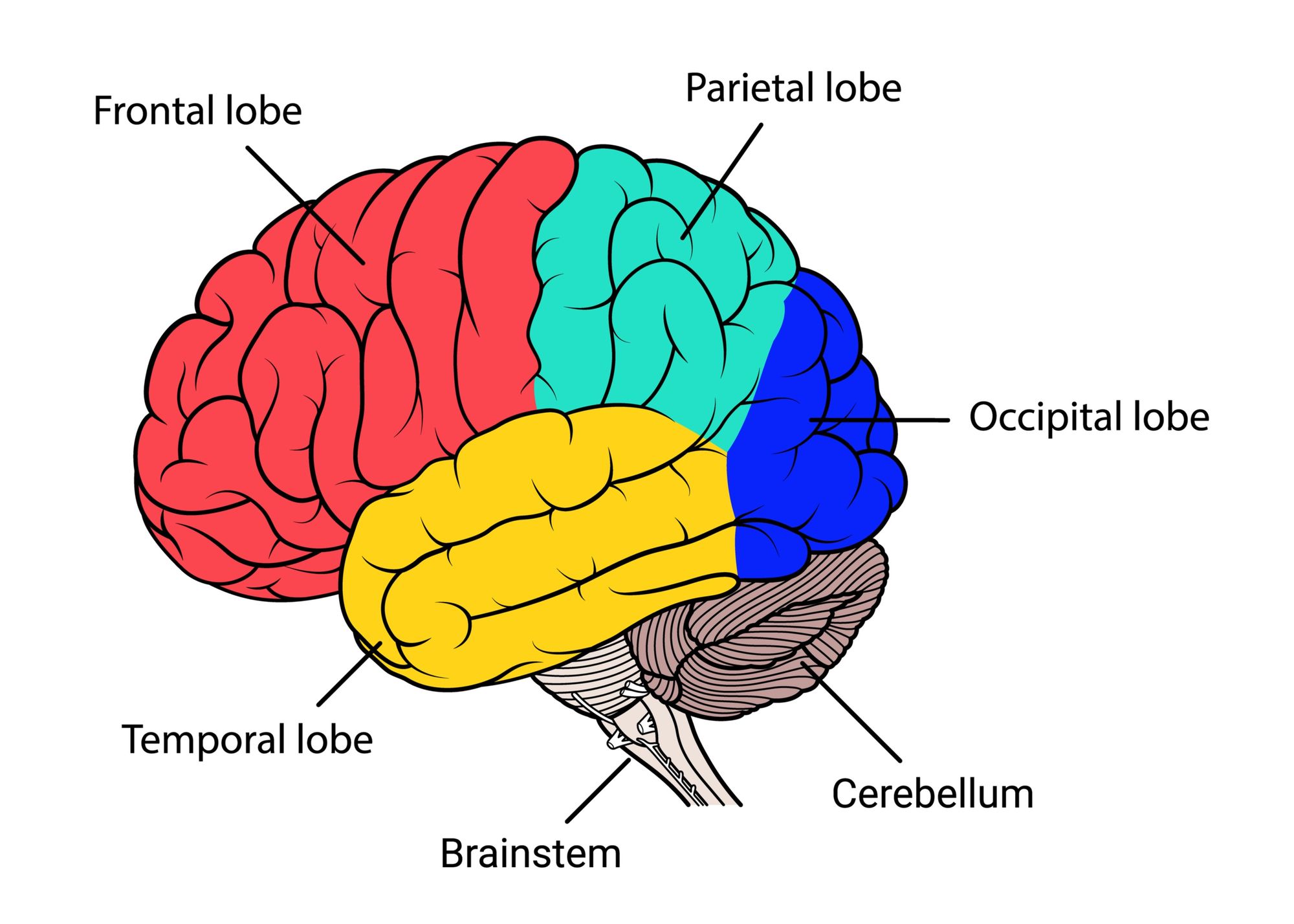
The brain diagram given below highlights the different lobes of the human brain. Where is the Brain located? The brain is enclosed within the skull, which provides frontal, lateral and dorsal protection. The skull consists of 22 bones, 14 of which form the facial bones and the remaining 8 form the cranial bones.

Related image Anatomia del cerebro humano, Partes del cerebro humano
The labeled human brain diagram contains labels for: The frontal lobe, parietal lobe, temporal lobe, occipital lobe, cerebellum, and brainstem. The diagram is available in 3 versions. The first version is color coded by section. The second version is the natural color of the human brain, and the third version is black and white.
.jpg)
Picture of human Brain Human Anatomy
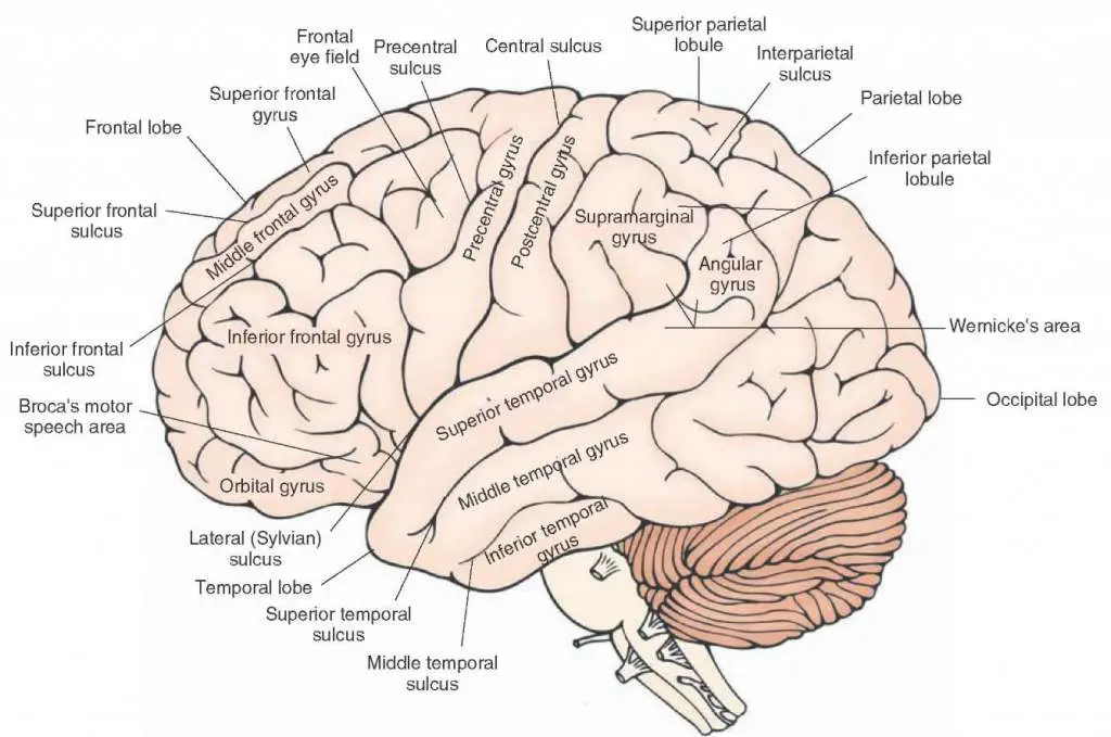
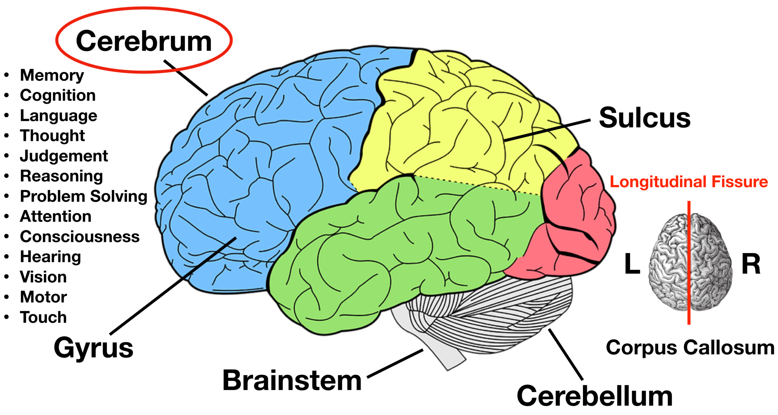
[Lateral views of the brain - labeled diagram]Looking at the brain from the lateral view we can see the frontal, temporal, parietal and occipital lobes. There are several important gyri and sulci that are visible from these two perspectives. The central sulcus separates the frontal from the parietal lobe (and the precentral gyrus from the.

Francisco's AP Macroeconomics Blog Psychology Unit 4Biological Basis
The cerebrum, also called the telencephalon, refers to the two cerebral hemispheres (right and left) which form the largest part of the brain. It sits mainly in the anterior and middle cranial fossae of the skull. The surface of the cerebrum is formed by an outer grey matter layer, which is thrown into a convoluted pattern of ridges and furrows.

Brain Jack Image กรกฎาคม 2013
Your brain contains billions of nerve cells arranged in patterns that coordinate thought, emotion, behavior, movement and sensation. A complicated highway system of nerves connects your brain to the rest of your body, so communication can occur in split seconds. Think about how fast you pull your hand back from a hot stove.

Human Brain Diagram Labeled, Unlabled, and Blank
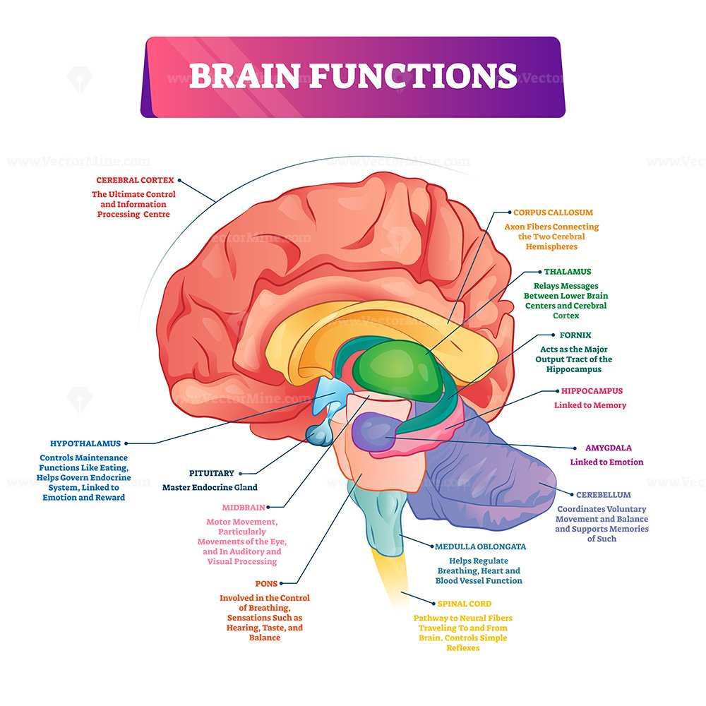
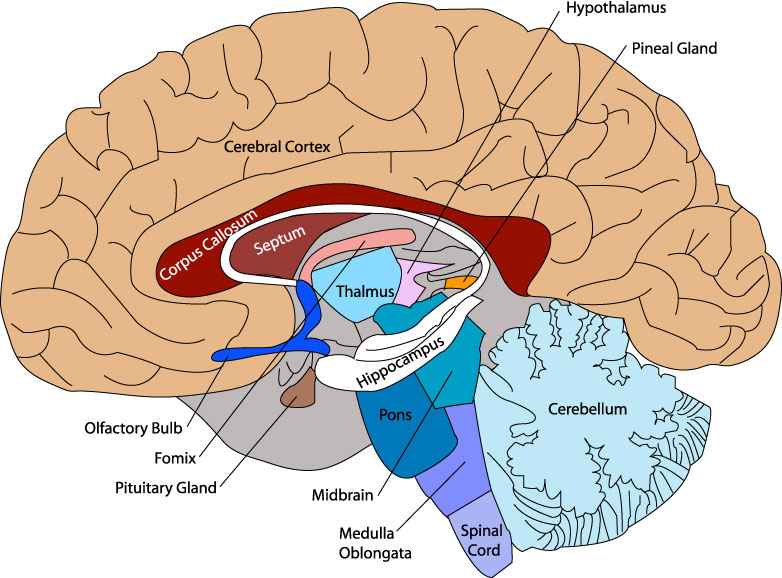
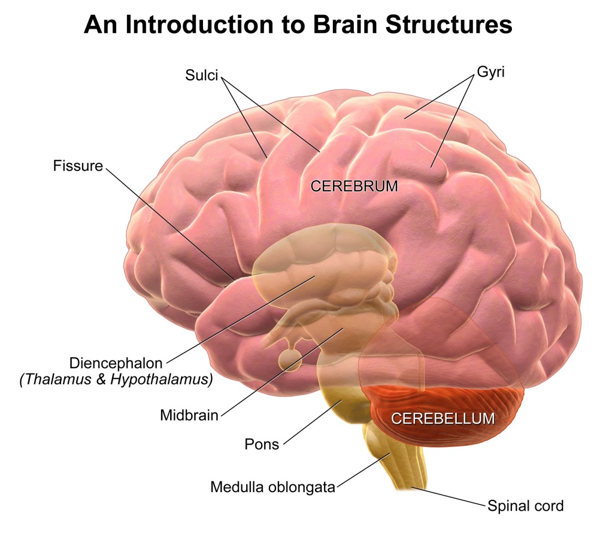
Brainstem: Anatomy: The brainstem is divided into 3 sections: the midbrain (mesencephalon), the pons (metencephalon), and the medulla oblongata (myelencephalon) Function: The brainstem is responsible for swallowing, breathing, vasomotor control (blood pressure) the senses - taste, smell, hearing, touch, sight, and controlling heartbeat.
Brain Jack Image Brain Diagram
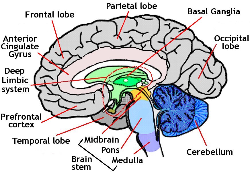
Anatomy of the Brain There are different ways of dividing the brain anatomically into regions. Let's use a common method and divide the brain into three main regions based on embryonic development: the forebrain, midbrain and hindbrain. Under these divisions:
Brain diagram
Figure 1. The brain has three main parts: the cerebrum, cerebellum, and brainstem. Cerebrum The cerebrum is the largest and most recognizable part of the brain. It consists of grey matter (the cerebral cortex ) and white matter at the center.

Colored And Labeled Human Brain Diagram Stock Illustration Download
The brain is made up of three main parts, which are the cerebrum, cerebellum, and brain stem. Each of these has a unique function and is made up of several parts as well. Keep reading to learn.
Unit 3 All About the Brain AP Psychology
The cerebral cortex is the part of the brain that is responsible for a number of complex functions including information processing, language, and memory. The Four Lobes
Parts of the Human Brain
Brain diagram. Use this interactive 3-D diagram to explore the brain. Anatomy and function. Cerebrum. The cerebrum is the largest part of the brain. It's divided into two halves, called.
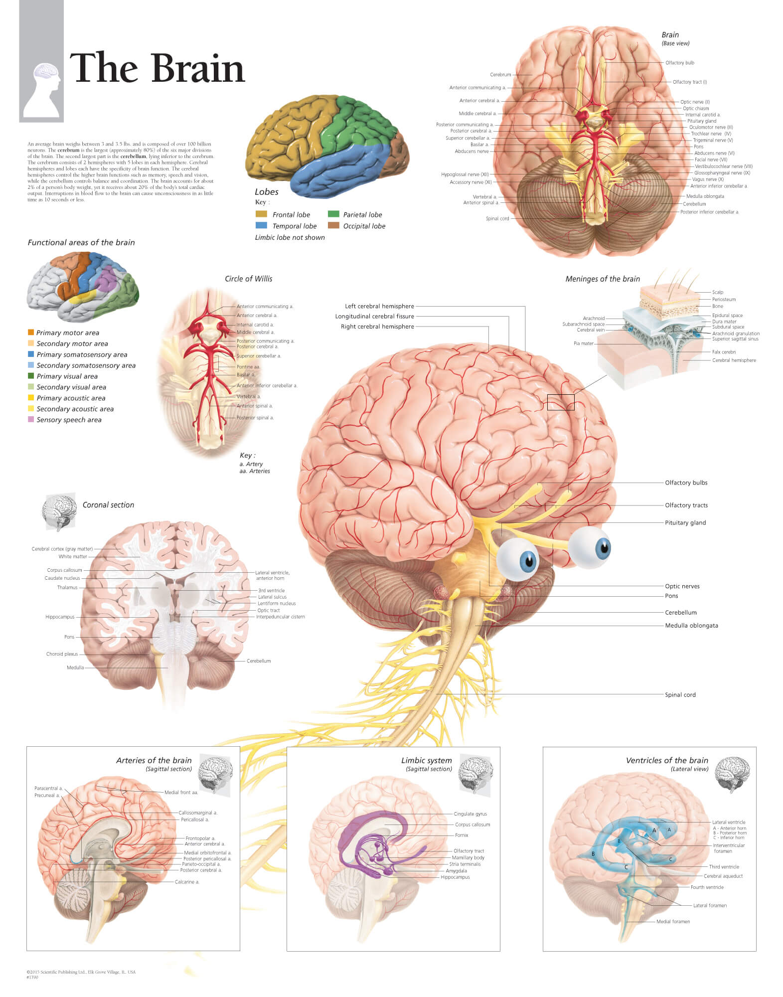
The Brain Scientific Publishing
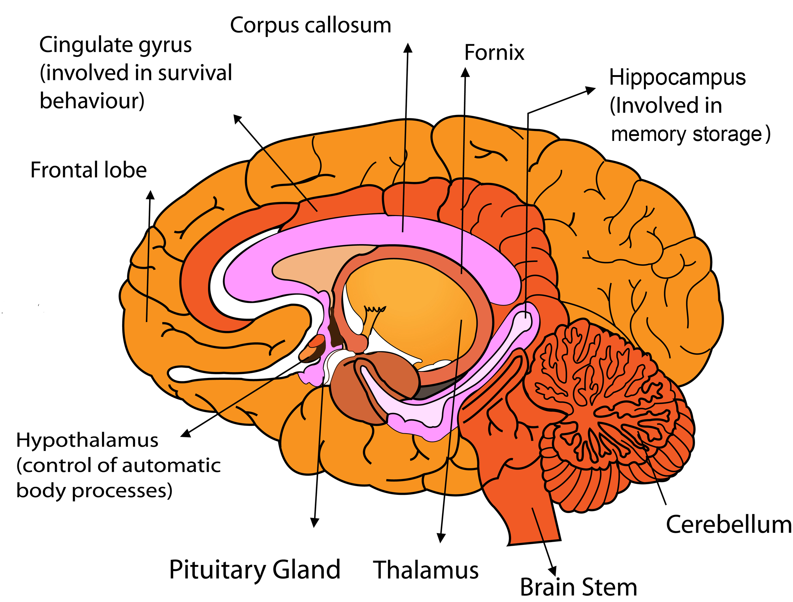
A topographical anatomy of the brain showing the different levels (encephalon, diencephalon, mesencephalon, metencephalon, pons and cerebellum, rhombencephalon and prosencephalon) as well as a diagram of the various cerebral lobes (frontal lobe, occipital, parietal, temporal, limbic and insular). Please note that the limbic lobe is functional.
Brain Images Labeled
The diagram of the brain is useful for both Class 10 and 12. It is one among the few topics having the highest weightage of marks and is frequently asked in the examinations. A well-labelled diagram of a human brain is given below for further reference. Structure And Function Of The Human Brain Parts Of The Human Brain

Image result for labeled diagram of the brain Brain diagram, Human
3D Brain. This interactive brain model is powered by the Wellcome Trust and developed by Matt Wimsatt and Jack Simpson; reviewed by John Morrison, Patrick Hof, and Edward Lein. Structure descriptions were written by Levi Gadye and Alexis Wnuk and Jane Roskams.
The Human Brain Facts, Anatomy, and Functions HubPages
How does the brain work? The brain sends and receives chemical and electrical signals throughout the body. Different signals control different processes, and your brain interprets each. Some make you feel tired, for example, while others make you feel pain.
PostStroke Dizziness How Vestibular Therapy Can Help
+ Show all Diagrams Diagrams are the perfect way to get orientated with a structure's detailed anatomy. Read on to see how we recommend using them. If you need some help with labeling the following diagrams, check out this video where we show you how to do it step-by-step: Labeled brain diagram