
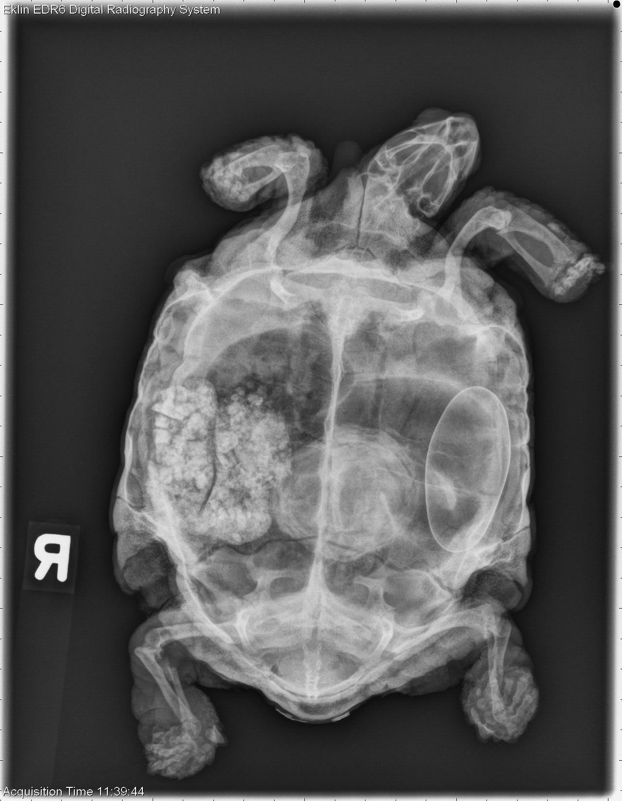
PREGNANT TORTOISE
Conscious turtles and tortoises have the ability to hold their extremities and head within or close to their shells. In the instance of fracture, sedation or anesthesia may be needed to relax the neck or extremity and isolate it, avoiding superimposi-tion with the shell, for complete evaluation. Restraint Techniques
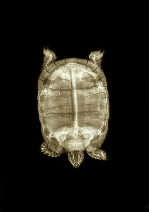
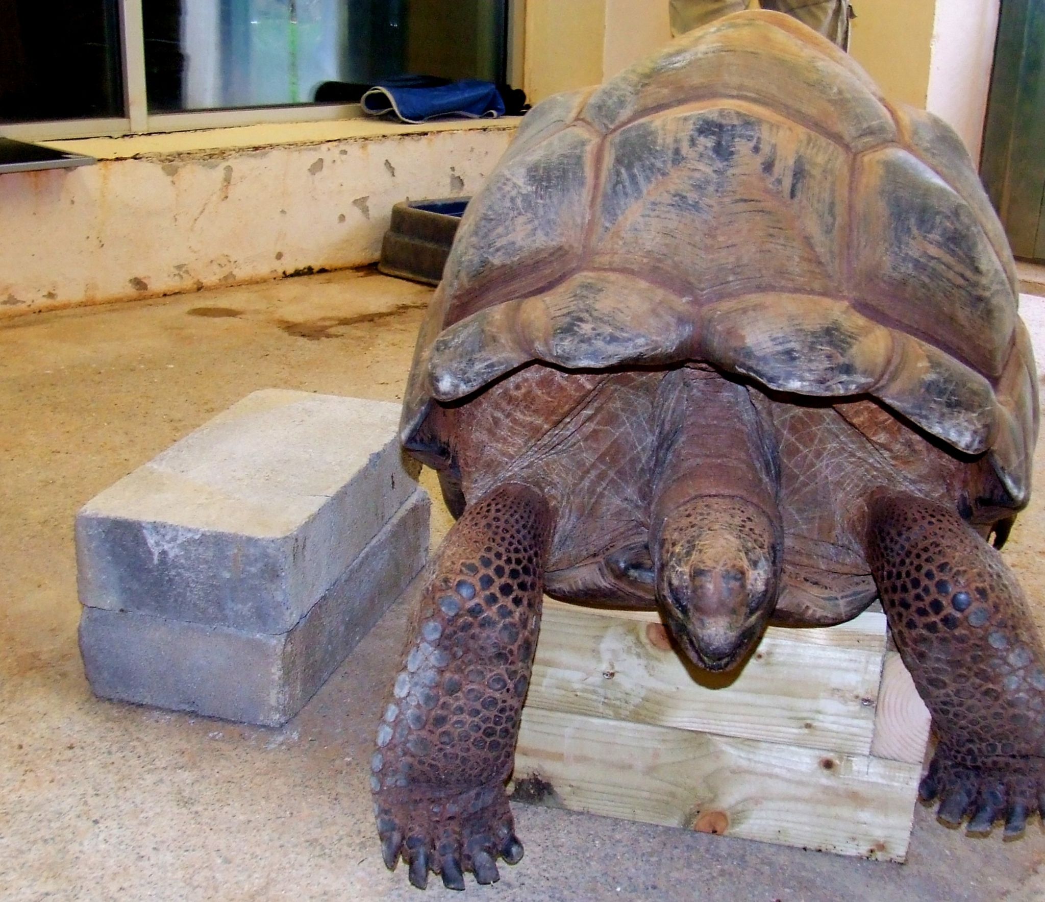
43 year old Desert Tortoise
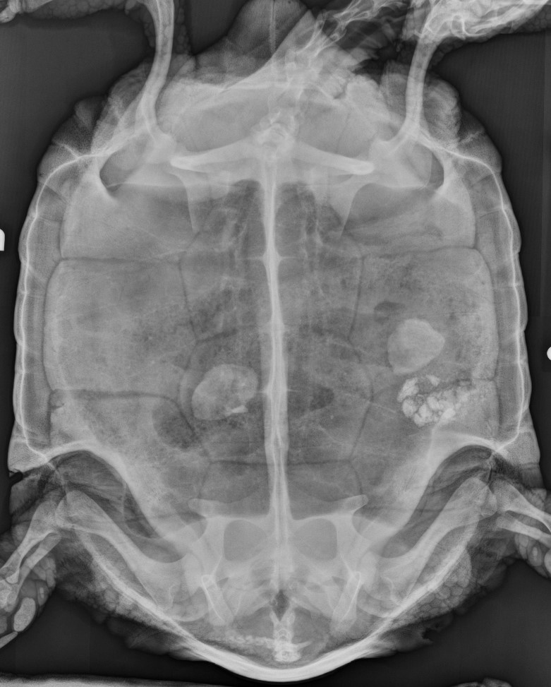
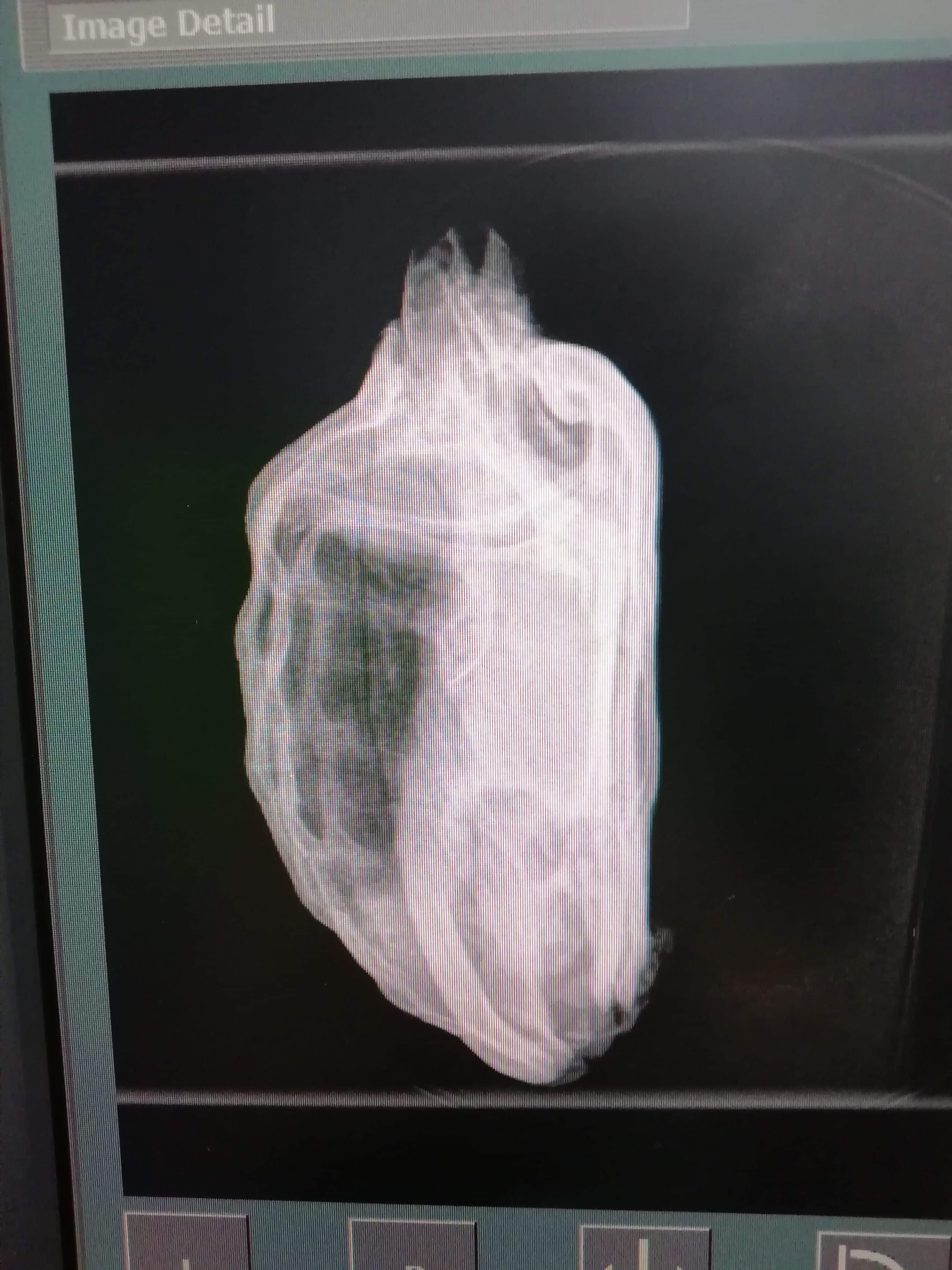
#1 tomatowide New Member Joined Feb 18, 2021 Messages 5 Location (City and/or State) Daegu, Korea unfortunately, my tortoises seems sick. So my family brought them to vet to check the status. And they sent some X-ray images to me. I wish the tortoises are fine, but are these really the proofs of pneumonia?

2006 December 11 ScienceRoll
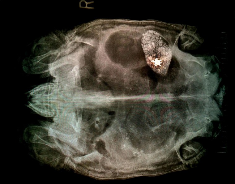
technique it need to knows the tortoise anatomy and be experienced at palpation [6,9]. Not every bladder stone can be found on palpation technique. The other more reliable method for diagnosing a bladder stone in a tortoise is to take an X-ray imaging. X-ray imaging result showing a white dense mass in the urinary bladder [4,6,9].

Turtle xray a photo on Flickriver
Stephen J. Divers , BVetMed, DACZM, DECZM, FRCVS, Department of Small Animal Medicine and Surgery, College of Veterinary Medicine, University of Georgia Reviewed/Revised Jun 2020 | Modified Oct 2022 Physical Examination Snakes Lizards Tortoises, Turtles, and Terrapins Anesthesia and Analgesia Preanesthetic Assessment and Stabilization

Xray shows sick tortoise swallowed turtle pendant YouTube
Introduction The main challenge when imaging reptiles is their vast species differences and thus thorough knowledge of the anatomy and physiology of the species is required in order to make diagnostic quality radiographs and to interpret them adequately.
Tortoise Under Xray Photograph by Photostockisrael Fine Art America
The Japan-led XRISM (X-ray Imaging and Spectroscopy Mission) observatory has released a first look at the unprecedented data it will collect when science operations begin later this year. The satellite's science team released a snapshot of a cluster of hundreds of galaxies and a spectrum of stellar wreckage in a neighboring galaxy, which gives scientists […]
XRay Reveals Turtle Pendant in Sick Tortoise's Belly
Today's Veterinary Practice | Peer-Reviewed Veterinary Journal
Exotics Team Treats 61YearOld Tortoise School of Veterinary Medicine
Radiography Correct positioning is important. Animals can be taped down, or radiographed through a box or bag if not sedated. Three views are typically required: Dorsoventral view - take care; a healthy animal can move very quickly off the table! Take exposure between expiration and inspiration.
Tortoise xray Case Gallery Diagnostic Imaging Vet Nurse
A juvenile tortoise with MBD can experience abnormal bone growth, consisting of curved bones, deformities, and thickened joints, leading to impaired movement, pain, and stiff walking.. allowing the professional to diagnose your tortoise's condition accurately. X-Ray - Radiographies are meant to investigate the bone structure to determine.
Giant tortoises prove Xray challenge at Paignton Zoo BBC News
No information was given on if the patient moved much during the x-ray proceedure, but the clarity of the x-ray helped this tortoise to a fast recovery. ABOVE: An X-ray showing the stone inside a tortoise bladder (BluePearl Veterinary Partners photo).

Tortoise under xray Stock Image C022/5261 Science Photo Library
Reptile Radiography Issue: November/December 2014 Danielle Mauragis CVT Clifford R. Berry DVM, DACVR Radiography of reptile patients is routinely used for evaluation of traumatic injuries and the gastrointestinal and reproductive tracts. A reptile radiography study typically includes lateral and dorsoventral views.

Pin on Reptiles, Amphibians, Tortoises
Cause The exact cause is unknown. Dehydration plays a significant factor in this disease. Many people believe that tortoises get all the water they need from their food. This is not true, they need to drink water also, so make sure fresh water is available all day. Periodic soaking is recommended.

xray of female turtle carrying eggs Vet Medicine, Veterinary Medicine
April 20, 2022 Turtle Radiography Here at the Wildlife Medical Clinic, we take digital radiographs, or x-rays, when we want to examine patients of all species and sizes. Radiographs are images of the body used to evaluate internal structures like organs and bones.

Turtle and Tortoise Diseases Crazy Plants Crazy Critters
Normal anatomy of chameleon Bearded dragon Frog (amphibian) distended with fluid Chuckwalla urinary bladder stone Chuckwalla urinary bladder stone This is the forearm and foot of a California Desert Tortoise (CDT) This tortoise is filled with eggs Can you guess the species just by looking at the x-ray?

Odd, eerie and cool Minnesota Zoo shares animal Xrays MPR News
The use of perspex or even cardboard boxes to constrain the reptile, particularly if they are some of the smaller lizards such as anoles and day geckos, is very useful, although minor reduction in the quality of the radiographs will occur.

Tiny sea turtle rescued with 158 pieces of plastic in gut
Radiographic Positioning and Technique for Reptiles. Read this imaging article by Jodi Nugent-Deal discussing proper positioning and techniques for turtles, tortoises, snakes, and lizards.