Lungs Respiratory System Coloring Page Work Sheet For Kids Coloring Home
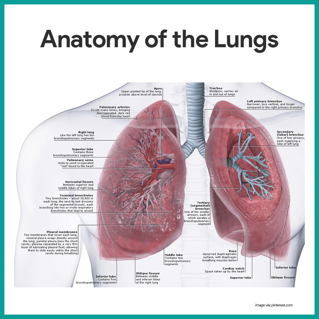
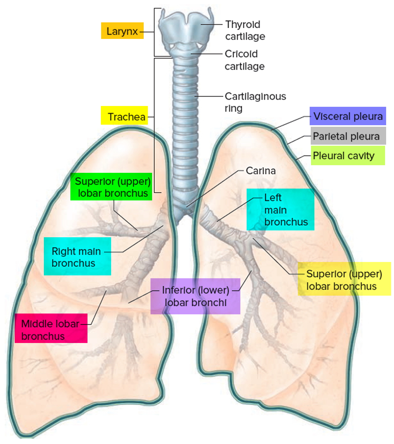
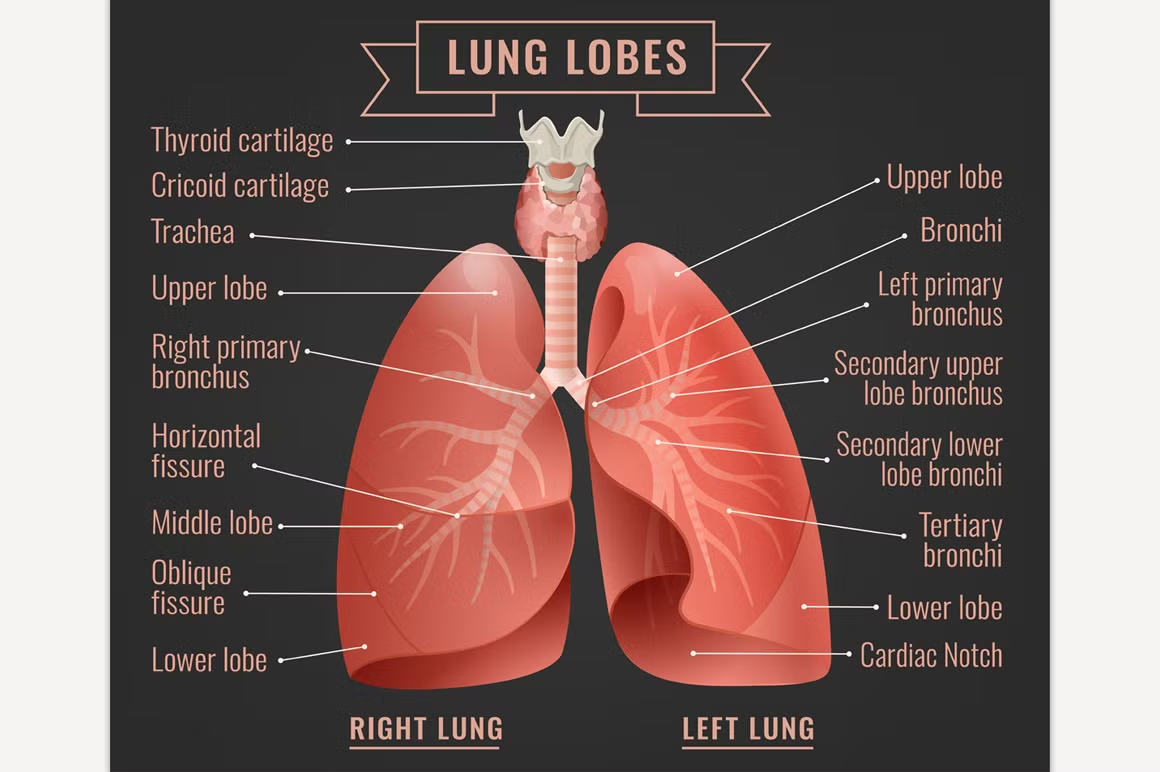
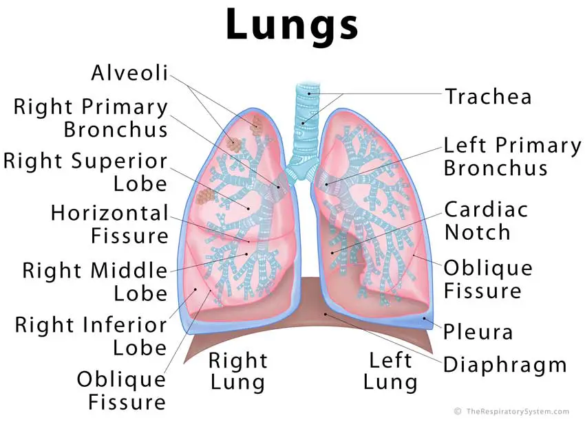
Functionally, the lung is divided into a series of bronchopulmonary segments. The bronchopulmonary segments are the largest subdivision of a lobe. They are separated from adjacent segments by connective tissue septa and are also surgically resectable. They are 10 bronchopulmonary segments in the left lung and 8-10 in the left lung [9].

Lung Diagrams Diagram Link
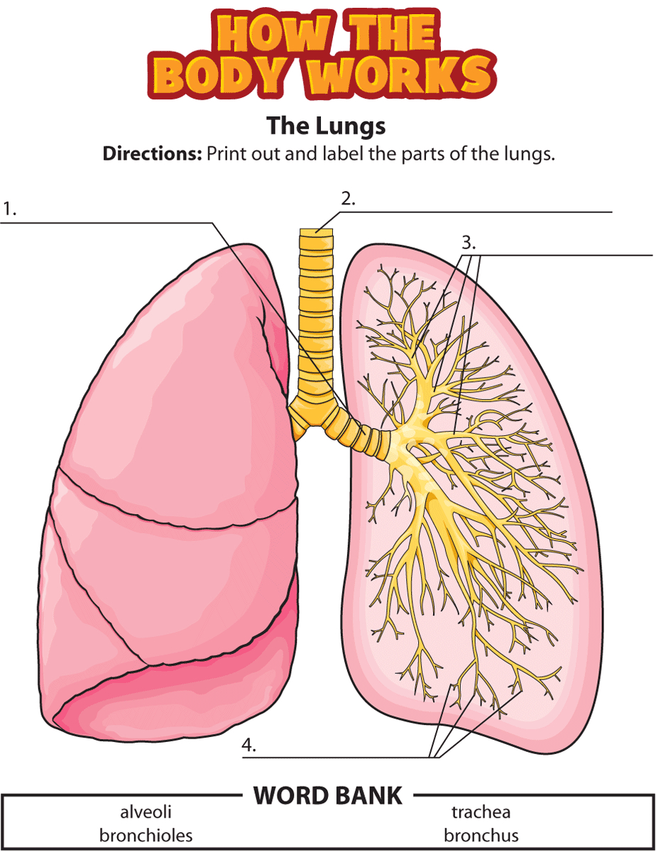
Label the Lungs by Kelsfied 1,852 plays 14 questions ~40 sec English 14p More 3 too few (you: not rated) Tries Unlimited [?] Last Played February 22, 2022 - 12:00 am There is a printable worksheet available for download here so you can take the quiz with pen and paper. Remaining 0 Correct 0 Wrong 0 Press play! 0% 0:00.0 Other Games of Interest
Lungs clipart labeled, Lungs labeled Transparent FREE for download on WebStockReview 2023
The lungs are the organs of respiration. They are located in the thorax, either side of the mediastinum. The function of the lungs is to oxygenate blood. They achieve this by bringing inspired air into close contact with oxygen-poor blood in the pulmonary capillaries.
Respiratory System Anatomy and Physiology Nurseslabs
The respiratory system, also called the pulmonary system, consists of several organs that function as a whole to oxygenate the body through the process of respiration (breathing). This process involves inhaling air and conducting it to the lungs where gas exchange occurs, in which oxygen is extracted from the air, and carbon dioxide expelled.
CrossFit Anatomy of the Lungs
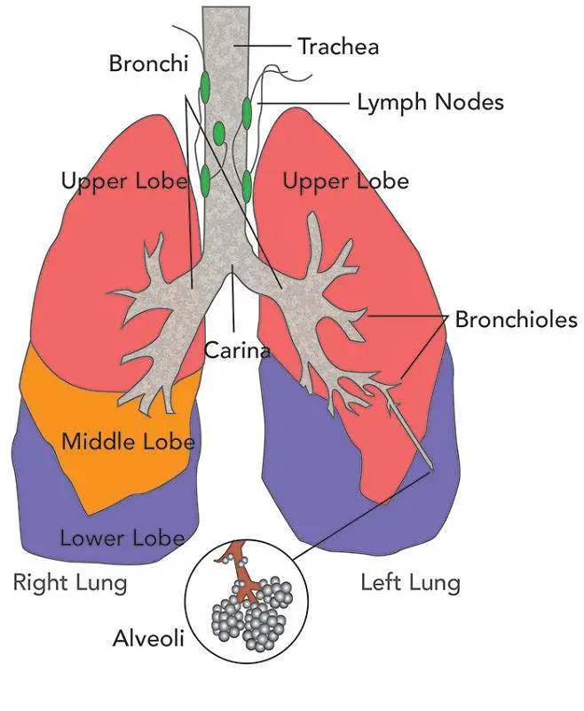
What is the Respiratory System? As defined above, the human respiratory system consists of a group of organs and tissues that help us to breathe. Aside from the lungs, there are also muscles and a vast network of blood vessels that facilitate the process of respiration. Also Read: Mechanism of Breathing Human Respiratory System Diagram
Lung Anatomy & Function Lung Nodule, Lung Disease and Lung Infection
Interactive for 5th Graders This online quiz is called Respiratory System Labeling Interactive. It was created by member teacherrojas and has 11 questions.
The lungs Lung cancer Macmillan Cancer Support
An ala is a cartilaginous structure that forms the lateral side of each naris (plural = nares), or nostril opening. The philtrum is the concave surface that connects the apex of the nose to the upper lip. Figure 22.3 Nose This illustration shows features of the external nose (top) and skeletal features of the nose (bottom).
Lungs clipart labeled, Lungs labeled Transparent FREE for download on WebStockReview 2023
Lungs: Together the lungs form one of the body's largest organs. They're responsible for providing oxygen to capillaries and exhaling carbon dioxide. Bronchi: The bronchi branch from the.

Medical Education Chart of Biology for Lungs Diagram. Vector illustration Ad , sponsored,
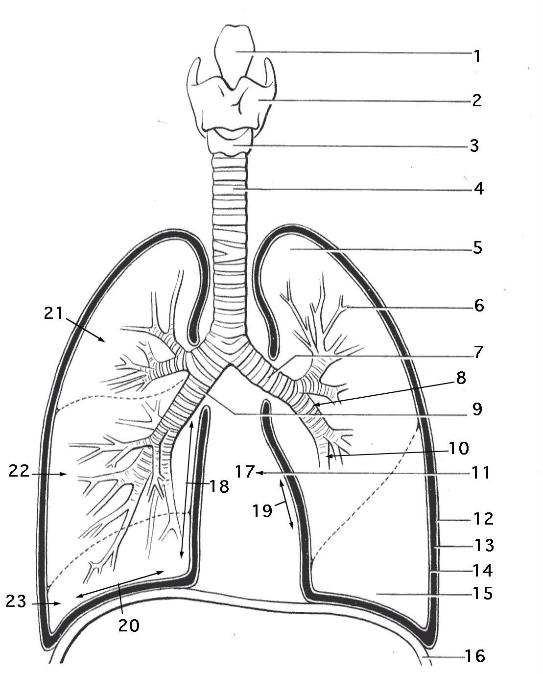
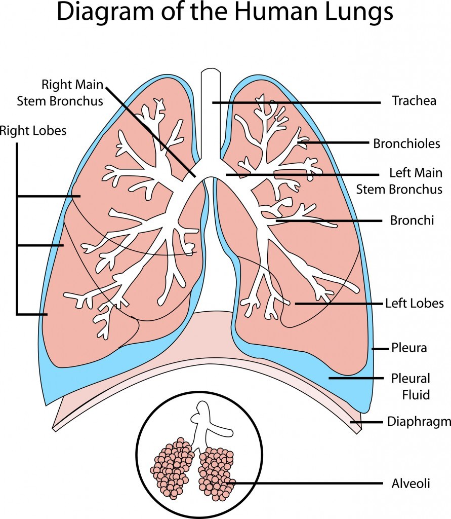
Labeled Diagram of the Human Lungs Lungs are an excellent example of how several tissues can be compactly arranged, yet providing a large surface area for gaseous exchange. The current article provides a labeled diagram of the human lungs as well as a description of the parts and their functions.
Human lungs infographic Education Illustrations Creative Market
Respiratory System Diseases. Common diseases of the respiratory system include: Asthma. Your airways narrow and make too much mucus. Bronchiectasis. Inflammation and infection make your bronchial.
A healthy lung has a pinkish appearance, and if you could see it outside the body, it would look
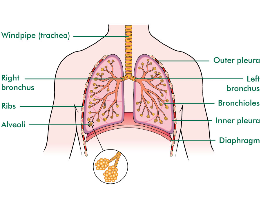
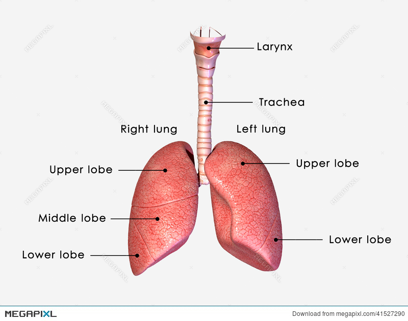
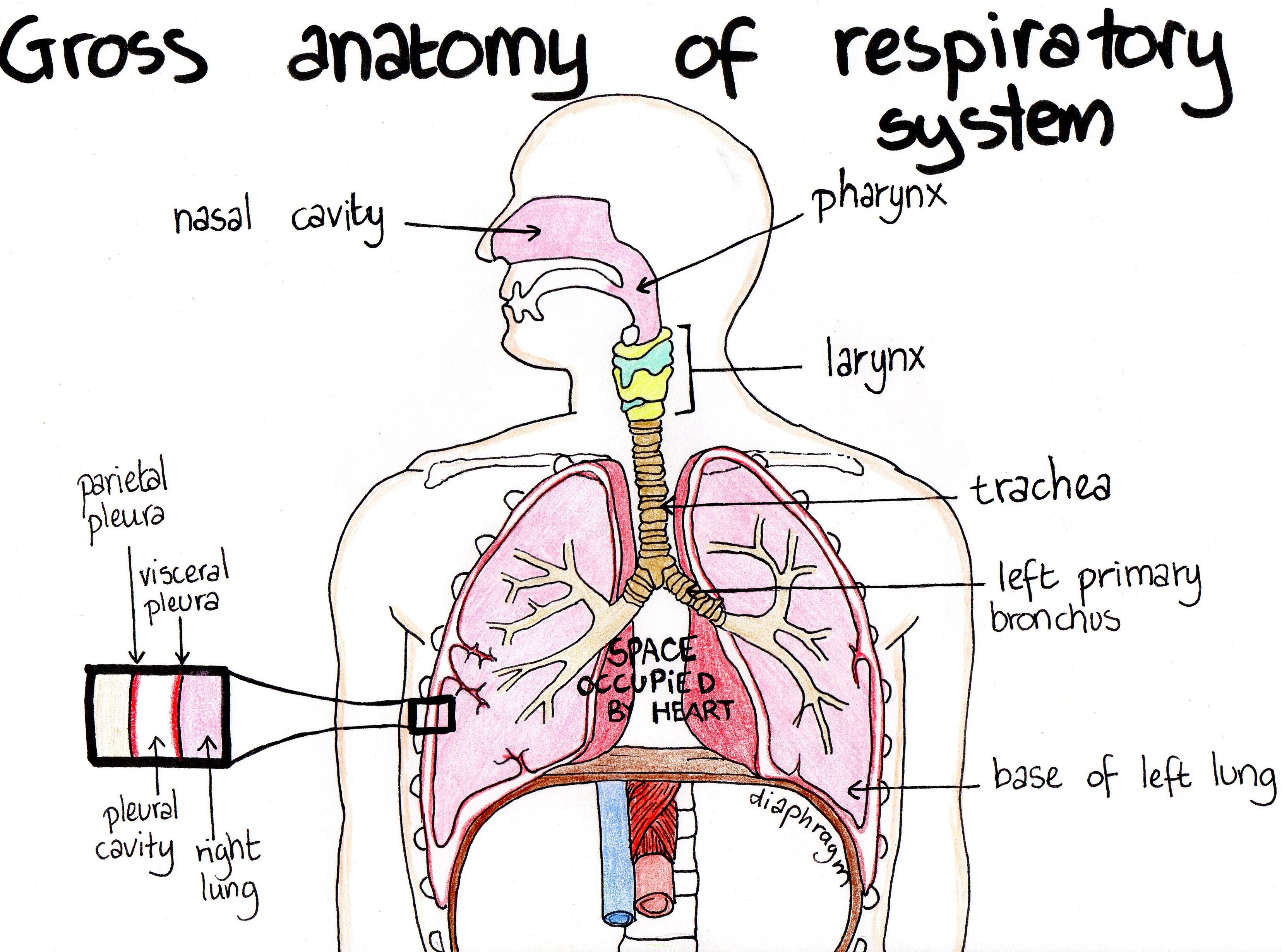
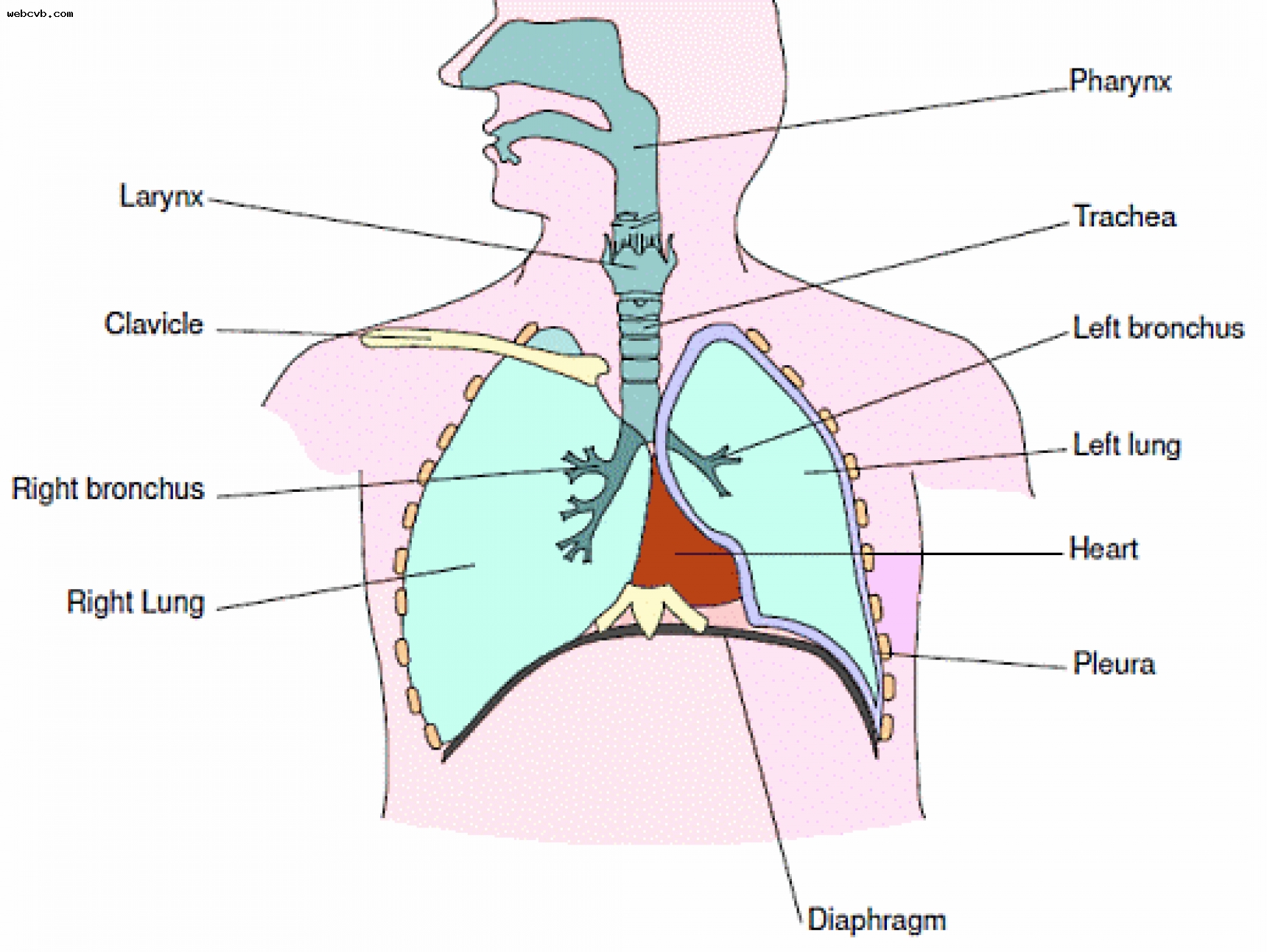
What are the Lungs? The lungs are respiratory organs - we use them to breathe. We breathe in order to get oxygen (O 2) and to get rid of carbon dioxide (CO 2 ). We breathe in through the nose or mouth. The air then goes through the larynx and the trachea (also called the windpipe) and into the lungs.
The Heart and Circulation Revision Cards in GCSE Biology
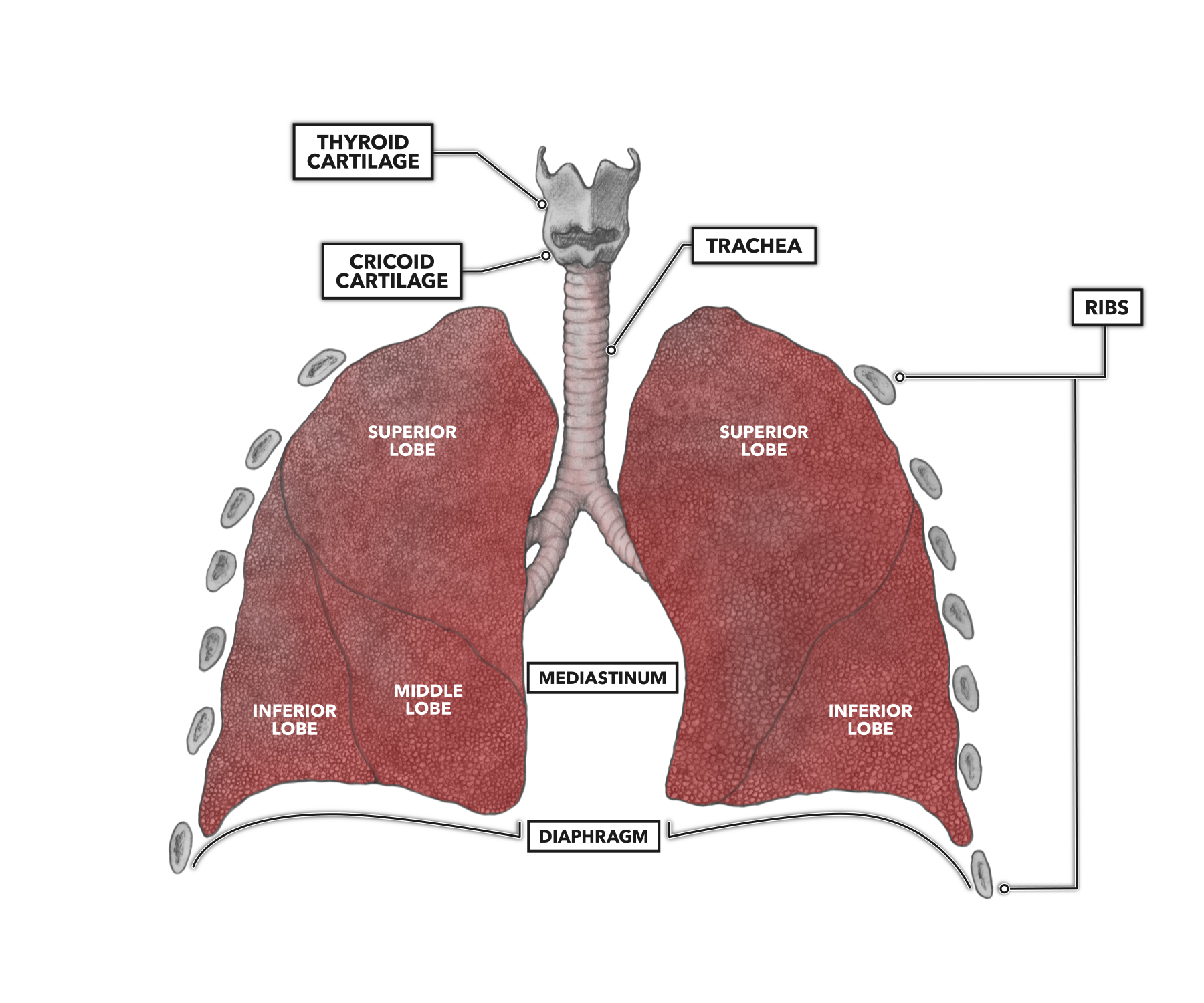
The lungs are pyramid-shaped, paired organs that are connected to the trachea by the right and left bronchi; on the inferior surface, the lungs are bordered by the diaphragm. The diaphragm is the flat, dome-shaped muscle located at the base of the lungs and thoracic cavity.
Respiratory System Medical Biology illustrated notes
The lungs are a major organ that is part of the respiratory system, taking in fresh air and getting rid of old, stale air. This mechanism of breathing also helps to allow you to talk. By taking in fresh air, the lungs are able to help oxygenate blood to be carried around your body. This is done by inhaling the air and bringing it in toward the.
Respiratory System Coloring Page Coloring Home Printable Worksheets On The Lungs Printable
Blood supply. The lungs have dual, parallel blood supply referred to as pulmonary and systemic circuits. The pulmonary circuit arises from the heart and brings deoxygenated blood to the lung for gas exchange. This circuit also takes oxygenated blood back to the heart to be redistributed to the rest of the body.
Lungs clipart respiratory system, Lungs respiratory system Transparent FREE for download on
The respiratory tract conveys air from the mouth and nose to the lungs, where oxygen and carbon dioxide are exchanged between the alveoli and the capillaries. Sagittal view of the human nasal cavity. The human gas-exchanging organ, the lung, is located in the thorax, where its delicate tissues are protected by the bony and muscular thoracic cage.

The lungs, trachea and bronchi, mediastinum and detail of chest wall and pleurae. Created in
Can you label the lungs? by birdb08 2,991 plays 9 questions ~30 sec English 9p 4 too few (you: not rated) Tries 9 [?] Last Played February 22, 2022 - 12:00 am There is a printable worksheet available for download here so you can take the quiz with pen and paper. From the quiz author Labeling the lungs. Remaining 0 Correct 0 Wrong 0 Press play! 0%