Unlabeled Dog Skeleton Diagram Data Diagram Medis
Dog Printouts. Read the definitions below, then label the dog external anatomy diagram. back - the part of the body between the loin and the withers. brisket - the chest of the dog. carpals - the wrist, the bones of the pastern joint. dewclaw - the tiny, useless, fifth claw - located on the inner part of the leg above the other toes.
Anatamation where Anatomy meets Animation Dog anatomy
Large laminated wall poster illustrating the anatomy of a dog skeleton, including the skull, teeth and limbs. £18.00. Inc VAT. £15.00. Exc VAT. Qty. Made in UK. Free UK Delivery on Orders over £50. Rated Excellent on Reviews.io.

Dog skeleton Dog skeleton, House training dogs, Dogs
This veterinary anatomical atlas includes selected labeling structures to help student to understand and discover animal anatomy (skeleton, bones, muscles, joints, viscera, respiratory system, cardiovascular system). Positional and directional terms, general terminology and anatomical orientation are also illustrated.
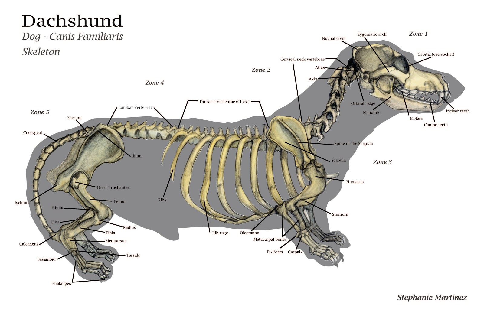
SM[art]inez November 2012
Forelimb Hindlimb Joints Bone types and parts of the dog skeleton Regarding bone types, the dog skeleton is made of three main types of bones: long, irregular (no particular shape) and flat bones In the big picture, the dog skeleton is made of two basic parts: axial and appendicular (limbs).

Print of Skeleton of a greyhound in 2020 Dog anatomy, Dog skeleton, Animal skeletons
The cat has a small coronoid fossa medial to the radial fossa that accommodates the coronoid process of the ulna during elbow joint flexion.; The cat has a supracondylar foramen near the medial condyle allowing the passage of the median nerve and brachial blood vessels.; There is an intermediate tubercle between the greater and lesser tubercles in the horse's intertubercular groove.

Dog Skeleton Anatomy ubicaciondepersonas.cdmx.gob.mx
Dog anatomy comprises the anatomical studies of the visible parts of the body of a domestic dog.Details of structures vary tremendously from breed to breed, more than in any other animal species, wild or domesticated, as dogs are highly variable in height and weight. The smallest known adult dog was a Yorkshire Terrier that stood only 6.3 cm (2.5 in) at the shoulder, 9.5 cm (3.7 in) in length.

Art of Lucia Dog study, and some life drawing
Xiphoid region (Cranial abdominal region) Zygomatic bone. Zygomatic gland. Zygomatic region. Radiographic anatomy: labeled images in the transverse plane of a healthy dog's whole body, using tomodensitometry. Introduction to the anatomy of the skull, thorax, abdomen, pelvic cavity, muscles and blood vessels: main anatomical structures identified.

Dog skeleton with major bone elements labeled (Davis, 1987, p. 54;... Download Scientific Diagram
In this module of the animal atlas vet-Anatomy is displayed the cross-sectional labeled anatomy canine thorax on a Computed Tomography (CT) and on 3D images of the thorax of the dog. CT images are available in 3 different planes (transverse, sagittal and dorsal) with two kinds of contrast (bones/lungs and soft tissues/mediastinum/vessels).
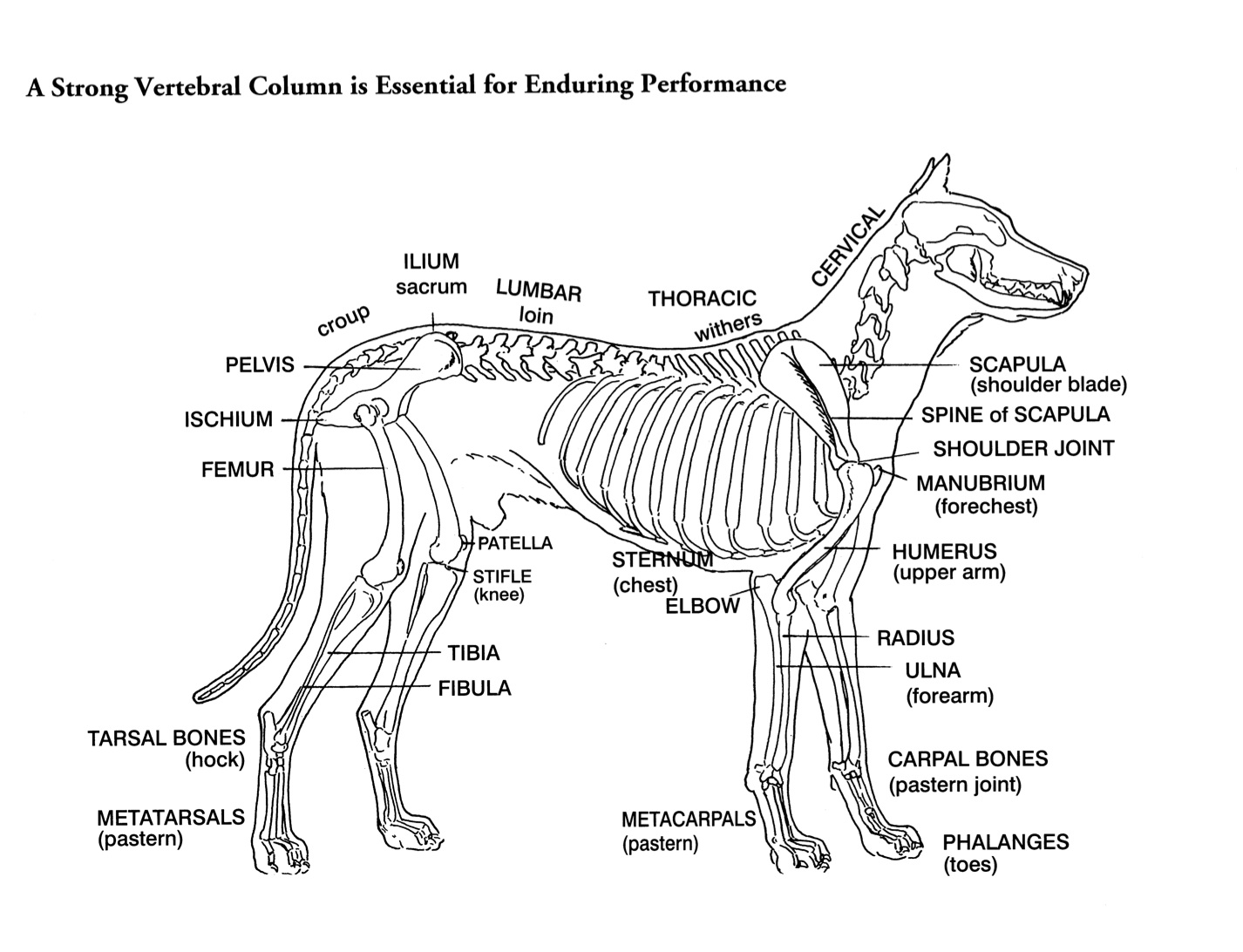
Helen King on Structure Evaluation Susan Garrett's Dog Training Blog
25/04/2023 31/12/2021 by Sonnet Poddar The dog skeleton anatomy consists of bones, cartilages, and ligaments. You will find two different parts of the dog skeleton - axial and appendicular. Here, I will show you all the bones from the axial and appendicular skeleton with their special osteological features.
Skeleton Worksheet Answers WikiEducator
This module of vet-Anatomy is a basic atlas of normal imaging anatomy of the dog on radiographs. 51 sampled x-ray images of healthy dogs performed by Susanne AEB Borofka (PhD - dipl. ECVDI, Utrecht, Netherland) were categorized topographically into seven chapters (head, vertebral column, thoracic limb, pelvic limb, larynx/pharynx, thorax and abdomen/pelvis).

Printable Anatomy Poster Dog Skeleton Canine Skeleton Etsy Australia
iStock Anatomy Of Dog Skeleton With Labeled Inner Bone Scheme Vector Illustration Stock Illustration - Download Image Now Download this Anatomy Of Dog Skeleton With Labeled Inner Bone Scheme Vector Illustration vector illustration now. And search more of iStock's library of royalty-free vector art that features Dog graphics available for quick and easy download.

Dog skeleton with major bone elements labeled (Davis, 1987, p. 54;... Download Scientific Diagram
It provides information about a dog's skeletal, reproductive, internal, and external anatomy, along with accompanying labeled diagrams. After mating, dogs experience something called a copulatory tie, wherein they remain in the coital position. The male dog dismounts the female at this time.
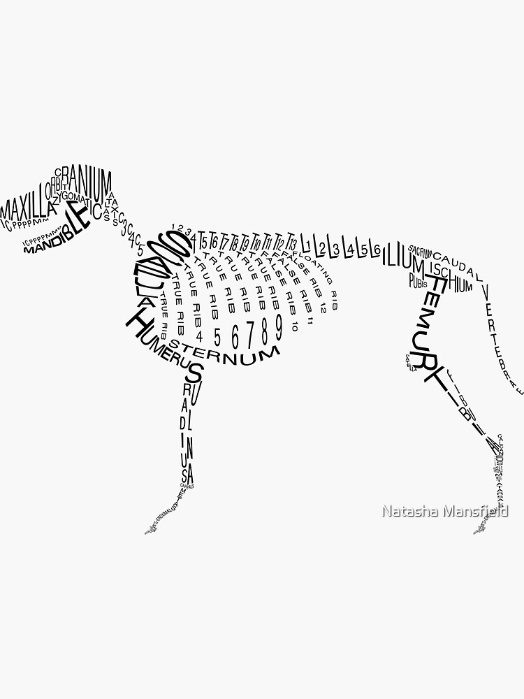
"Typographic Dog Skeleton" Sticker for Sale by howlinglights Redbubble
ISSN 2534-5087. This veterinary anatomy module of the dog contains 218 illustrations dedicated to the canine osteology anatomy. Here are presented scientific illustrations of the canine skeleton, with the main dog's bones and its structures displayed from different anatomical standard views (cranial, caudal, lateral, medial, dorsal, palmar..).

Resin Halloween Dog Skeleton Holidae Fun & Games
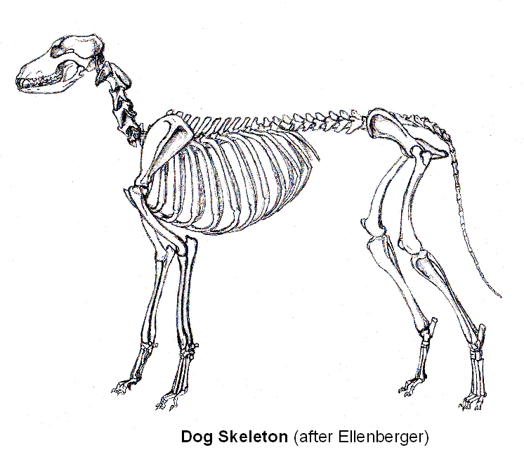
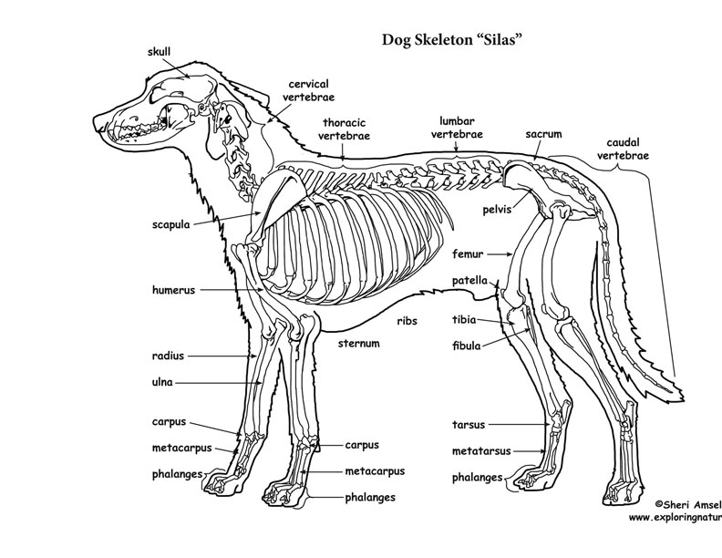
The skeleton is composed of the hard tissues of the body, and its primary functions are to support the body, to provide a system of levers used in locomotion, to protect the soft organs of the body, and to produce red blood cells (hematopoiesis). A dog's skeleton is formed so the dog can run fast, hunt and chase.

Labeled atlas of anatomy illustrations of the dog Bones Skeletal system Molecular Shapes
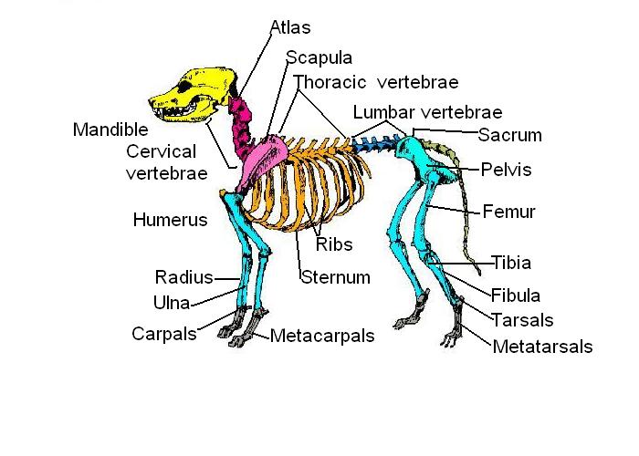
Image Skeleton of a dog Skeleton of a dog: carnivorous domestic mammal raised to perform various tasks for humans. Skull: bony case of the brain. Cervical vertebrae: bones of the neck. Thoracic vertebrae: the bones forming the dorsal part of the thoracic cage. Lumbar vertebrae: the bones of the lumbar region of the back.
Dog Skeletal Anatomy
This veterinary anatomy module contains 608 illustrations on the canine myology. Here are presented scientific illustrations of the canine muscles and skeleton from different anatomical standard views (lateral, medial, cranial, caudal, dorsal, ventral / palmar.). Some fascias, tendons, ligaments, joints were labeled.