
Vitals & Anatomy Horse Side Vet Guide Horse anatomy, Horses, Horses and dogs
The joint connecting the upper front leg (arm) and the forearm. Allows for bending and flexing of the front leg during movement. Ergot: A small, horn-like growth found on the back of the fetlock joint. Vestigial structure with no known function in modern horses. Face: The front of the horse's head, including the forehead, eyes, and muzzle.

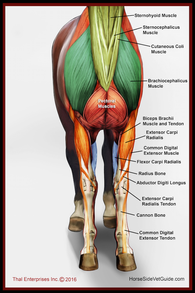
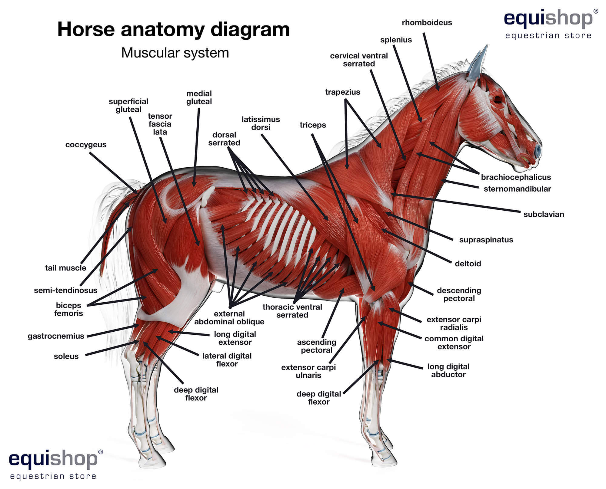
Horse Anatomy the Muscles by COOKEcakes on DeviantArt
Anatomy Equine leg anatomy comes with some specific terms. Here are some of the key parts you'll want to learn, starting at the top of the legs and working our way down: Stifle - Found on the hind legs only, the stifle is equivalent to the human knee joint. Located between the femur and the tiba, the stifle is below and behind the flank swirl.

Forever Horses Anatomy of the Equine Forleg
The elbow is the joint of the front leg where the leg meets the body. Ergot. The ergot is a callous, horn-like growth that may appear on one or all of the fetlock joints, along the backside. They are believed to be remnants of vestigial toes their ancestors had.. That's a lot of information for basic horse anatomy. However, this knowledge.

Leg anatomy Horse health, Horse care, Horse anatomy
1. Poll The poll is a boney protrusion located right behind the horse's ears, where the bridle begins. It's the highest point on the horse's head and may be more pronounced on some breeds than others. The poll's many nerve endings make it a spot that's prone to tension.

hip joint diagram muscles
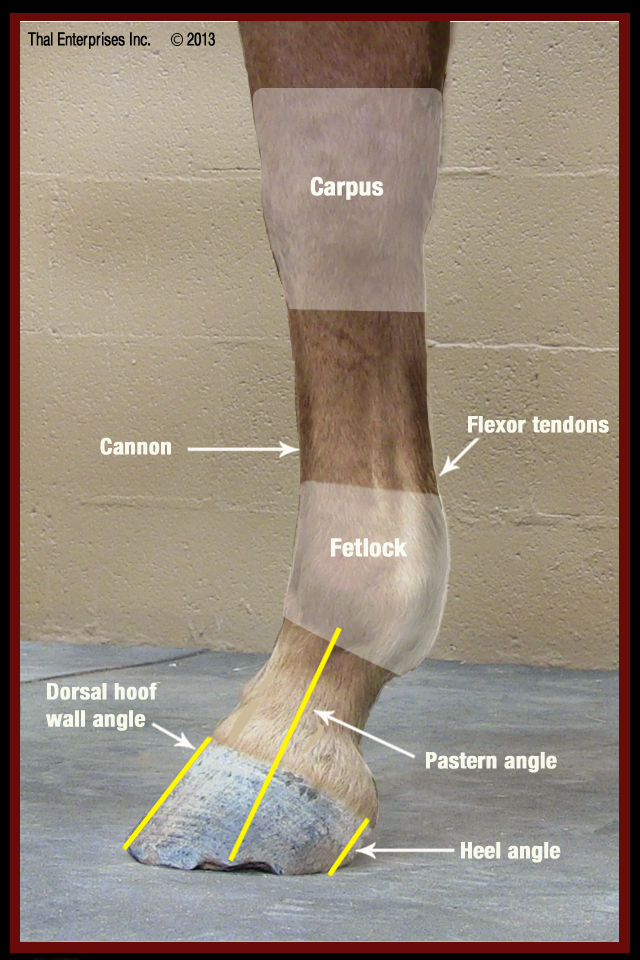
The digital flex-or tendons, which run down the back of the leg, support the leg's "suspension" and allow the hoof to flex and absorb shock. Meanwhile, the ligaments provide the necessary tension to maintain posture and balance. Why Care About Horse Leg Anatomy? Understanding the anatomy of a horse's leg is crucial for several reasons. It.

horse leg structure Google Search Horse anatomy, Horse health, Equestrian outfits
The best-known form of communication through the legs is probably the somewhat strange-looking resting position of horses, which includes a certain posture of one hind leg: In the relief position, one of the hind legs is placed on the front edge of the hoof, with the hoof joint (see anatomy) bending forward. Thus the leg no longer takes a load.
Horse Leg Anatomy Form and Function EquiMed Horse Health Matters
In horses, the front leg anatomy starts high at the shoulder joint. The shoulder joint in a horse has a limited range of motion, not being able to kick out to the side of their body, but well- developed and trained horses can gain incredible reach and lift in their stride.

Pin by Izabella Gowdy on Horse Anatomy Horse anatomy, Horse sculpture, Vet medicine
The front legs of a horse have parts owners and riders must learn to talk with trainers and vets. Learn about horse anatomy and parts of a horse's front legs from a ranch owner in.
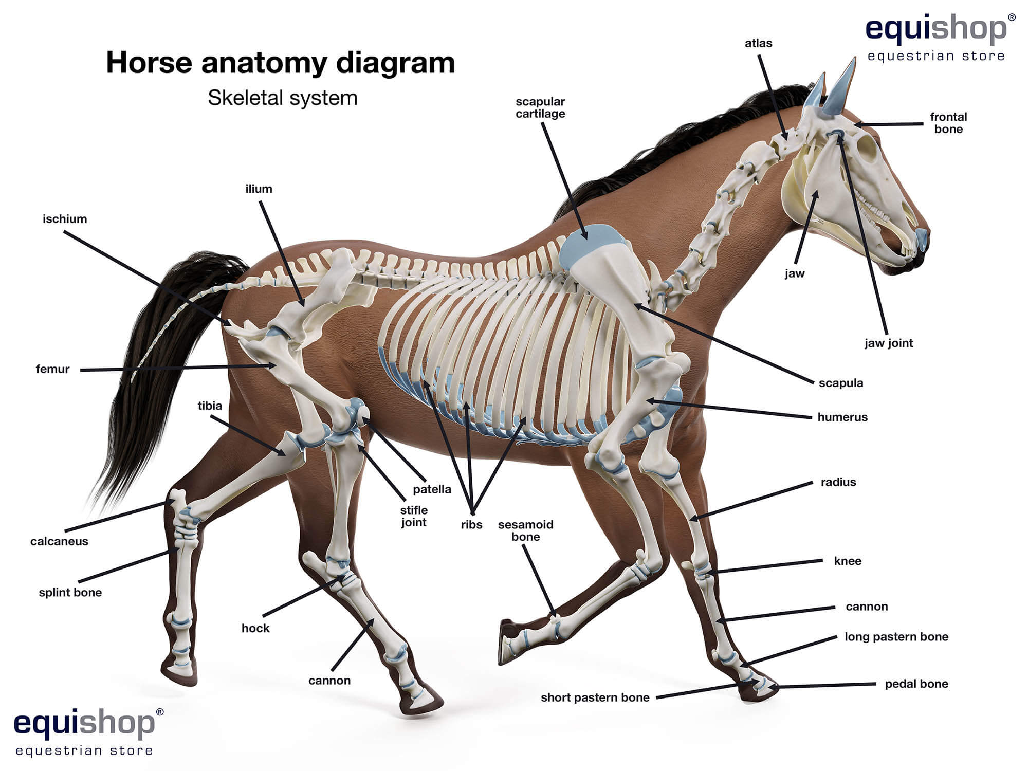
Horse anatomy diagrams of horse body parts Equestrian Shop
Anatomy of horse necks Types of horse necks - shapes and setting Horse barrel, or the trunk: Horse withers - the highest point How should the proper horse's chest look Types of horse chests Horse flank - a sensitive point Horse spine - or horse's back Types of horse backs Front and back limbs: Front limbs - function and build
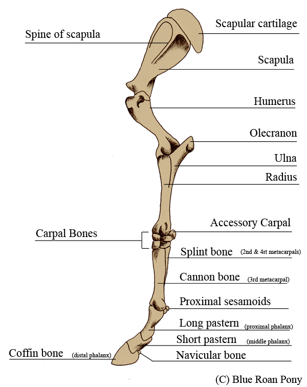
Forever Horses Anatomy of the Equine Forleg
The front legs of the horse carry approximately 60 percent of the weight of the horse and are constantly subject to lameness with approximately 95 percent of lameness occurring from the knee down, with the foot being the site of most problems.
Horse Anatomy Front Leg Anatomical Charts & Posters
Forms a broad forehead with a short bottom part and long top half of the head, typically seen in racing and breed horses. Dished - concave Broad forehead, large nostrils and eyes, and a concave line between the nose and small falcate ears. This look is mostly seen in Arab horses, including half-Arabs. Roman nose
Horse Anatomy Reference Vitals & Anatomy upyourbutthealing
Robert C. McClure Department of Veterinary Anatomy College of Veterinary Medicine A horse's hoof is composed of the wall, sole and frog. The wall is simply that part of the hoof that is visible when the horse is standing. It covers the front and sides of the third phalanx, or coffin bone.

Horse Leg Bones Diagram 187 Horse Anatomy Illustrations Royalty Free Vector Graphics Clip Art
The horse leg anatomy in the rear includes the bones of the pelvis (the ilium, ischium, and pubic bones), femur, tibia, fibula, metatarsus, and phalanxes. It also includes the joints of the hip, stifles, hock, fetlock, pastern, and coffin. Hind limbs The top part of the hind limbs consists of three fused bones, called the ileum, ischium, and pubis.

Pin by Amy Lynn on EVT Horses, Horse therapy, Horse anatomy
The limbs of the horse are structures made of dozens of bones, joints, muscles, tendons, and ligaments that support the weight of the equine body.
Anatomía del caballo diagramas de las partes del cuerpo del caballo Tienda Ecuestre
Toe - The front portion of the hoof that faces toward the head. Heel Bulbs - The two bulbous structures on the back of the hoof underneath the pastern. Ergot - A small, keratin growth on the back and underside of the horse's fetlock. Typically, this is protected or covered by hair, but can easily be located.

Equine Lower Limb From Front Horse anatomy, Anatomy, Horse health
Neck: The portion of the horse's body that is between the head and shoulders. Shoulder: The upper portion of the horse's front leg. Withers: The bony ridge at the base of the neck between the shoulder blades. This ridge is created by the top portion of the thoracic vertebrae. Horses are measured at the withers.