
Pictures Of The Animal Cells ANIMALSD
Diagram Of Animal Cell Animal cells are eukaryotic cells that contain a membrane-bound nucleus. They are different from plant cells in that they do contain cell walls and chloroplast. The animal cell diagram is widely asked in Class 10 and 12 examinations and is beneficial to understand the structure and functions of an animal.
Structure of cell Cell structure and functions, Class 8
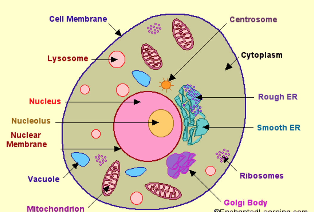
Looking for a Labeled Diagram of an Animal Cell? You Found It! What's inside an animal cell? Animal Cells are made up of a number of unique parts, including: Cell Membrane Nucleus Nuclear Membrane Centrosome Lysosome Cytoplasm Golgi Apparatus And more! Create Your Own Animal Cell Diagram - Printable Animal Cell Worksheets and More!

What is a cell? Facts
The shape of a typical animal cell varies widely from being flat, oval to rod-shaped, while others assume shapes such as curved, spherical, concave, and rectangular. An animal cell ranges in size from 10 to 30 µm. Under the microscope, an animal cell shows many different parts called organelles, that work together to keep the cell functional.

What Is An Animal Cell? Facts, Pictures & Info For Kids & Students.
1 Draw a simple circle or oval for the cell membrane. The cell membrane of an animal cell is not a perfect circle. You can make the circle misshapen or oblong. The important part is that it does not have any sharp edges. [1] Also know that the membrane is not a rigid cell wall like in plant cells.

South Pontotoc Biology Plant and Animal Cell Diagrams
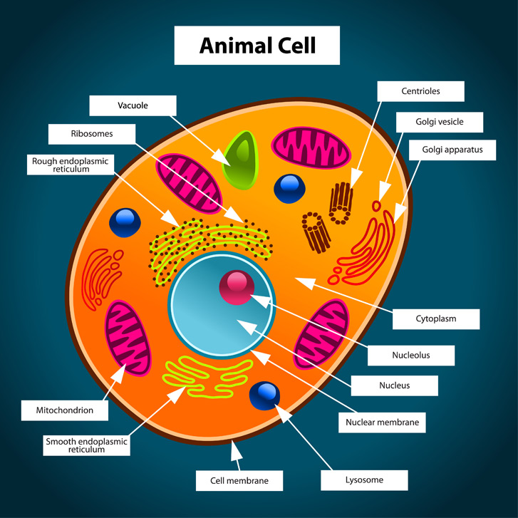
Labeled Animal Cell Diagram Blank Animal Cell Diagram Worksheet The third and fourth diagrams are animal cell diagram worksheets. Quiz yourself by filling in the blanks. Unlabeled Animal Cell Diagram Finally, an unlabeled version of the diagram is included at the bottom of the page, in color and black and white.
Discovery and Structure of Cells Biology Visionlearning
Home / Biology Detailed guide on Animal Cell and its parts (with labelled diagrams) By Editorial Team August 20, 2022 Introduction Animal cells are eukaryotic cells, mostly multicellular containing cytoplasm and membrane-bounded organelles enclosed within the plasma membrane.
Animal Cell Diagram CBSE Class Notes Online Classnotes123
In this guide Revise Video Audio Test Animal cells Almost all animals and plants are made up of cells. Animal cells have a basic structure. Below the basic structure is shown in the same.
Animal Cell Structure Carlson Stock Art
Definition Animal cells are the basic unit of life in organisms of the kingdom Animalia. They are eukaryotic cells, meaning that they have a true nucleus and specialized structures called organelles that carry out different functions.
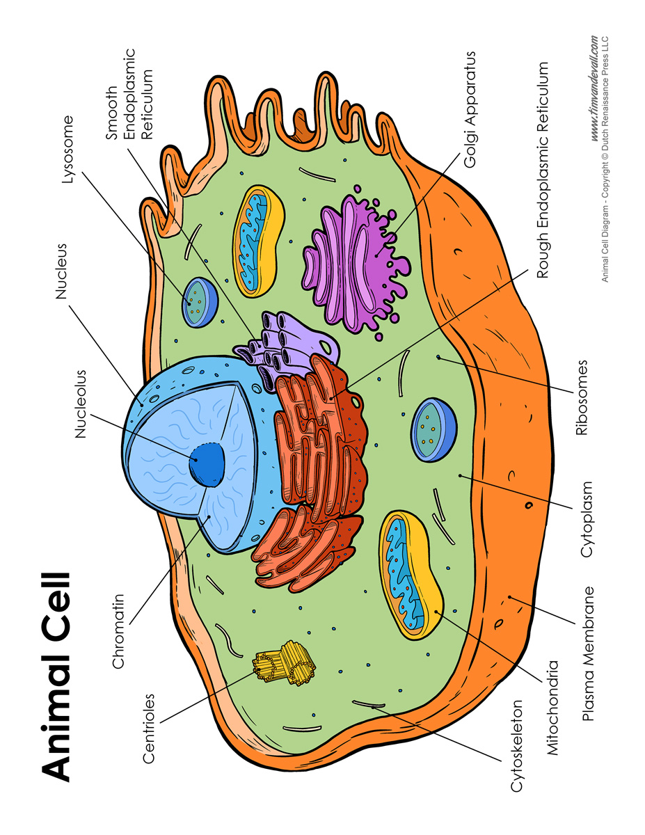
Animal Cell Diagram Labeled Tim van de Vall
A Labeled Diagram of the Animal Cell and its Organelles There are two types of cells - Prokaryotic and Eucaryotic. Eukaryotic cells are larger, more complex, and have evolved more recently than prokaryotes. Where, prokaryotes are just bacteria and archaea, eukaryotes are literally everything else.

The Structure and Functions of an Animal Cell
Well Labelled Diagram of Animal Cell Biology Study Material Biology top 10 Important Topics Biology Syllabus Biology Question Papers Book online demo NCERT Solutions NCERT Notes NCERT Important Question Difference Between Weather and Climate Scientific Names of Animals and Plants Bacterial Diseases in Humans Enzymes MCQs MCQs on Greenhouse Effect

Animal Cell Definition Structure Parts Functions Labeled Diagram Riset
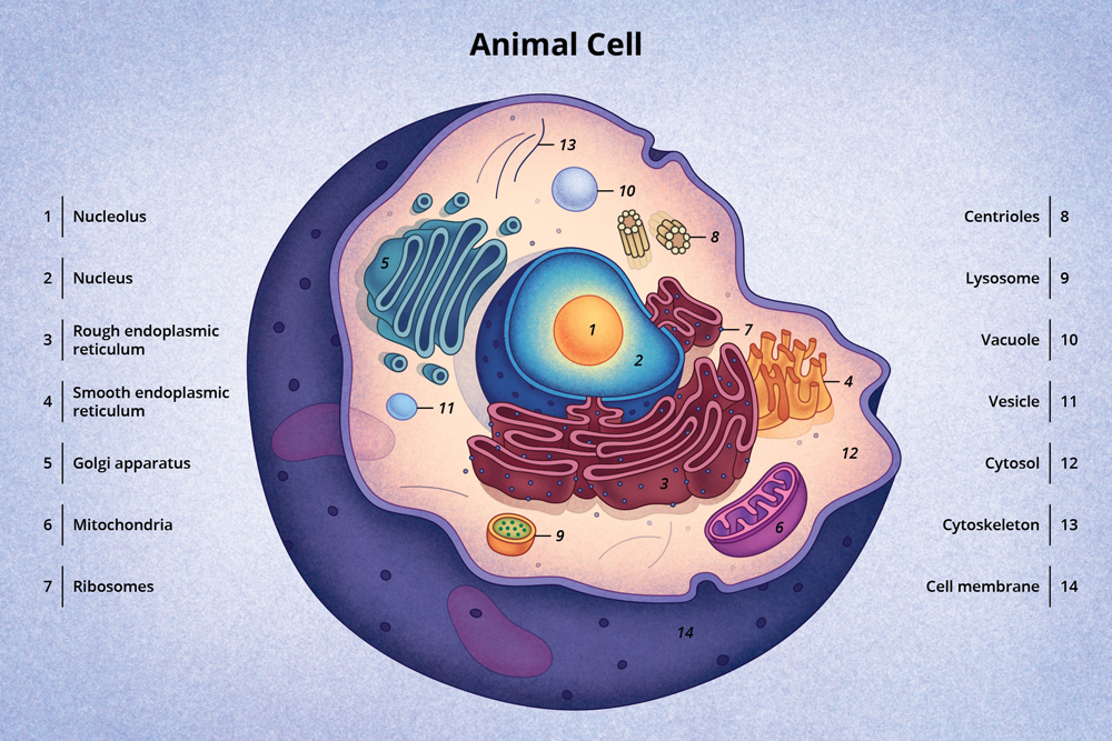
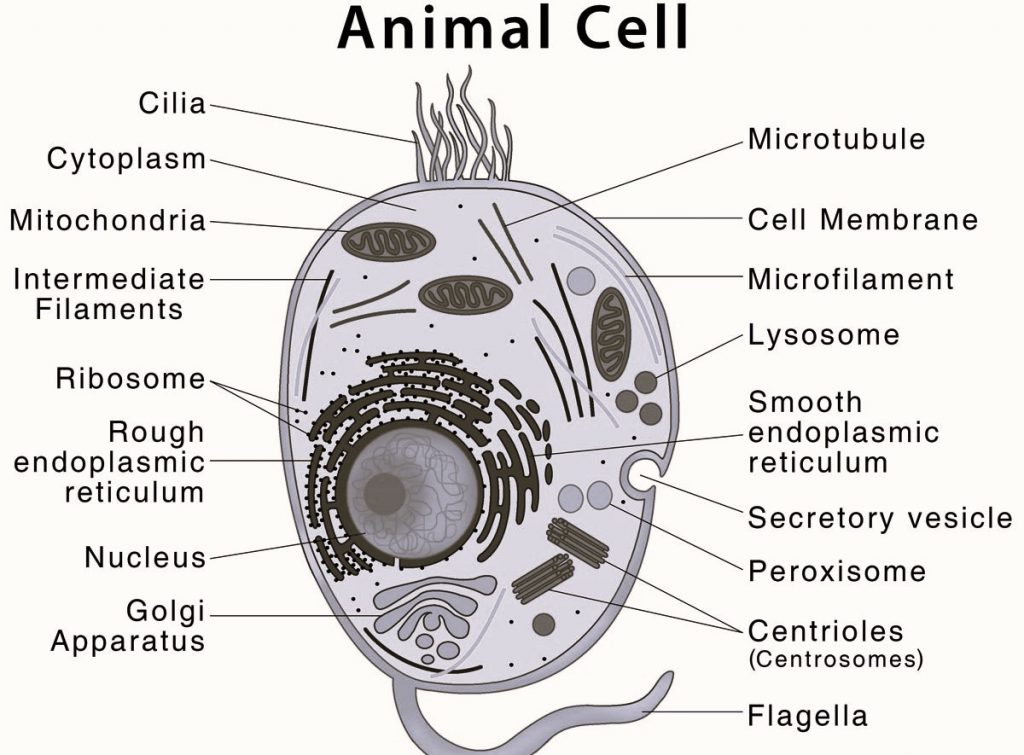
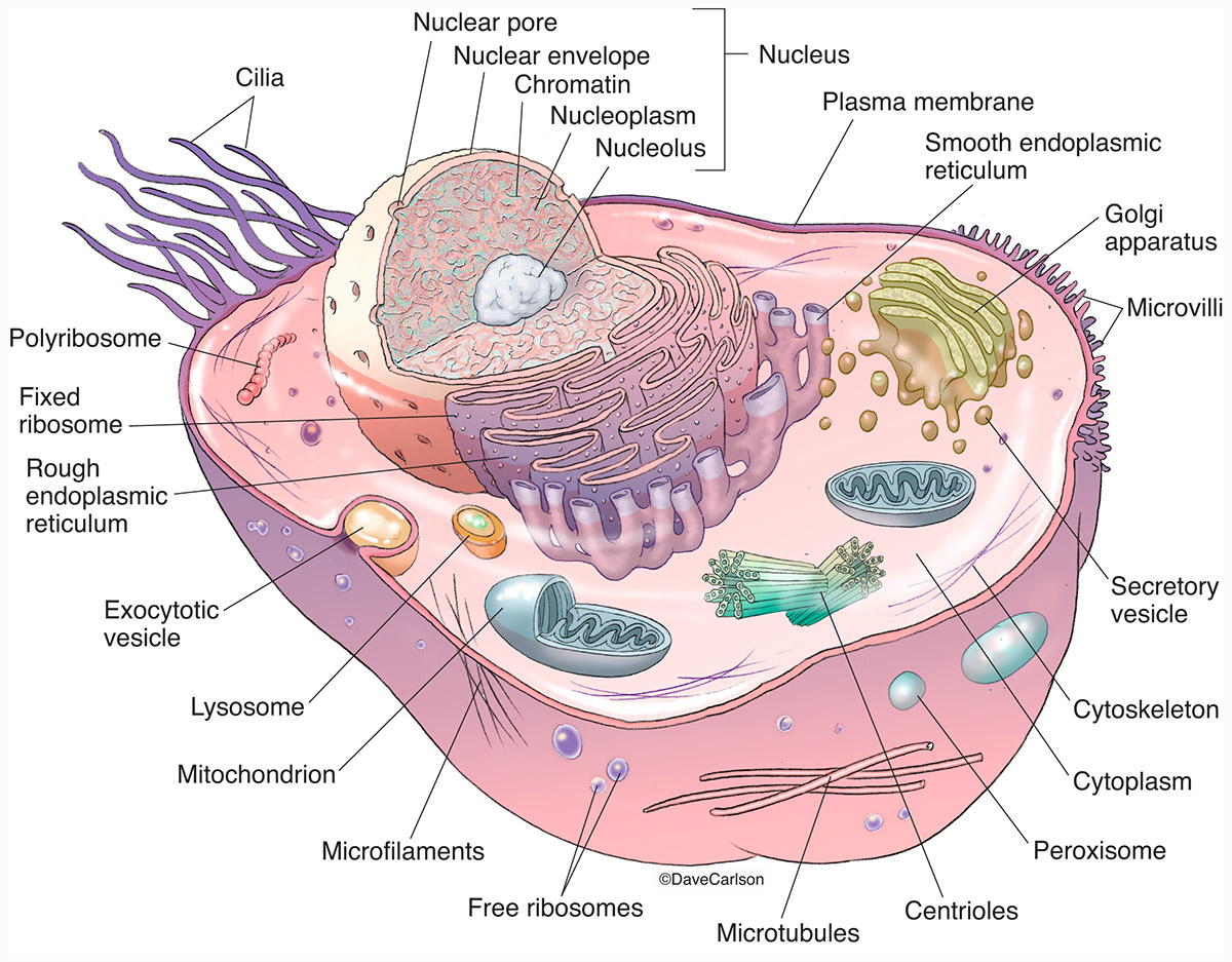
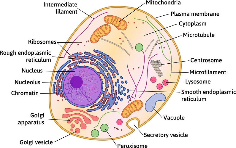
Overview of Animal Cells Animal cell size and shape Animal Cell Types Animal Cell Diagram and Structure (cross section of an animal cell) List of Animal cell organelles 1. Plasma membrane (Cell membrane) 2. Nucleus 3. Cytoplasm 4. Mitochondria 5. Ribosomes 6. Endoplasmic Reticulum (ER) 7. Golgi apparatus (Golgi bodies/Golgi complex) 8. Lysosomes 9.
Animal Cell The Definitive Guide Biology Dictionary
Animal Cell - Diagram, Organelles, and Characteristics This entry was posted on May 9, 2023 by Anne Helmenstine (updated on December 26, 2023) An animal cell lacks a cell wall or chloroplasts. Its outer coating is a semipermeable cell membrane. Animal cells are the fundamental units of life in protozoa and multicellular animals.
Animal Cell Free printable to label +
A flattened, layered, sac-like organelle that looks like a stack of pancakes and is located near the nucleus. It produces the membranes that surround the lysosomes. The Golgi body packages proteins and carbohydrates into membrane-bound vesicles for "export" from the cell. Lysosome (Cell Vesicles)
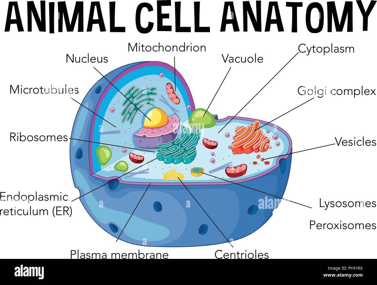
Diagram of animal cell anatomy illustration Stock Vector Image & Art Alamy
Key points: All cells have a cell membrane that separates the inside and the outside of the cell, and controls what goes in and comes out. The cell membrane surrounds a cell's cytoplasm, which is a jelly-like substance containing the cell's parts. Cells contain parts called organelles. Each organelle carries out a specific function in the cell.

Animal Cell Anatomy Anatomy Diagram Source Riset
Parts of Animal cell diagram . The Cell Organelles are membrane-bound and present within the cells. There are various organelles present within the cell and are classified into three categories based on the presence or absence of a membrane. Listed below are the Cell Organelles of an animal cell along with their functions.

The animal cell diagram. Vector illustration on white Etsy in 2021 Animal cell, Cell diagram
Learn about the structure and function of animal cells, the basic unit of life in animals. Explore the various organelles and their roles in maintaining homeostasis.. Labeled Diagram. By Go Life Science Posted on December 20, 2022 October 17, 2023. An animal cell is a type of cell that is characteristic of animals and is present in all.