
Respiratory System With Label Drawing at GetDrawings Free download
The respiratory system includes the organs, tissues, and muscles that help you breathe. It helps distribute oxygen throughout your body while filtering out carbon dioxide and other waste products.
Respiratory System With Label Drawing at GetDrawings Free download
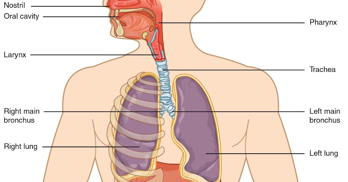
The respiratory tract conveys air from the mouth and nose to the lungs, where oxygen and carbon dioxide are exchanged between the alveoli and the capillaries. Sagittal view of the human nasal cavity. The human gas-exchanging organ, the lung, is located in the thorax, where its delicate tissues are protected by the bony and muscular thoracic cage.
RESPIRATORY SYSTEM
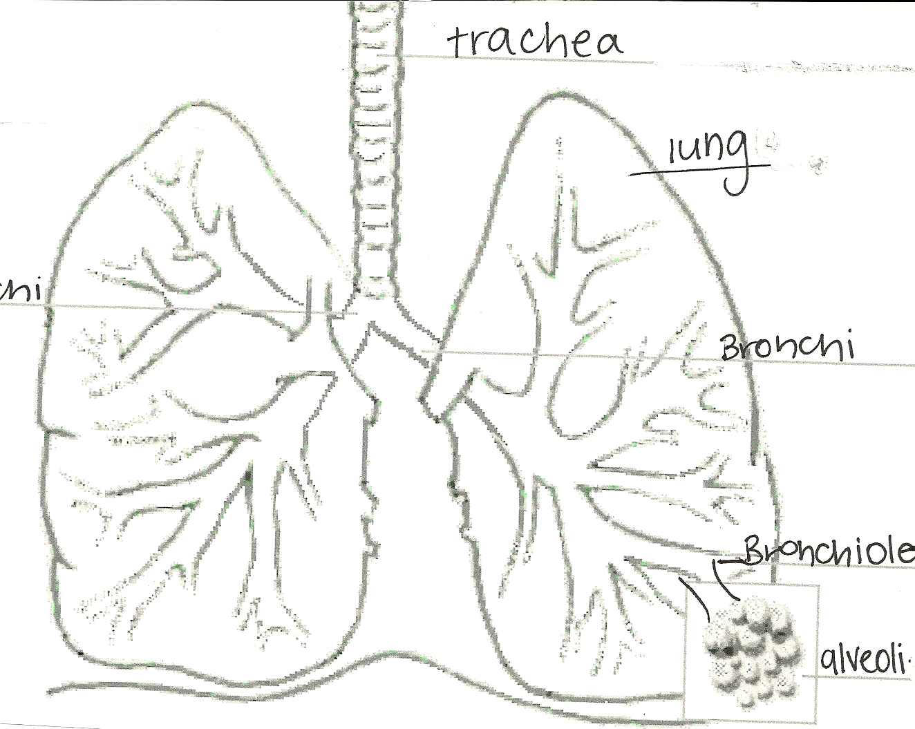
Respiratory system diagram Lower respiratory tract organs. Trachea: Also known as the windpipe this is the tube that carries air from the throat into the lungs. It ranges from 20-25mm in diameter and 10-16cm in length. The inner membrane of the trachea is covered in tiny hairs called cilia, which catch particles of dust that we can then remove.

C.1. Introduction to the Respiratory System
The respiratory system is the network of organs and tissues that help you breathe. It includes your airways, lungs and blood vessels. The muscles that power your lungs are also part of the respiratory system. These parts work together to move oxygen throughout the body and clean out waste gases like carbon dioxide. Advertisement.

The Respiratory Zone Of The Respiratory System Respiratory System No Labels Transparent PNG
Osmosis High-Yield Notes. This Osmosis High-Yield Note provides an overview of Anatomy and Physiology of the Respiratory System essentials. All Osmosis Notes are clearly laid-out and contain striking images, tables, and diagrams to help visual learners understand complex topics quickly and efficiently. Find more information about Anatomy and.
CLASS BLOG BIO 202 Respiratory System Worksheet
You are free: to share - to copy, distribute and transmit the work; to remix - to adapt the work; Under the following conditions: attribution - You must give appropriate credit, provide a link to the license, and indicate if changes were made. You may do so in any reasonable manner, but not in any way that suggests the licensor endorses you or your use.
Respiratory System With Label Drawing at GetDrawings Free download
Alveoli are connected to their neighbors by alveolar pores, which help maintain equal air pressure throughout the alveoli and lung ( Figure 22.11 ). Figure 22.11 Structures of the Respiratory Zone (a) The alveolus is responsible for gas exchange. (b) A micrograph shows the alveolar structures within lung tissue.

The Respiratory System Anatomy Sketch
The respiratory system is made up of the organs included in the exchange of oxygen and carbon dioxide. These are the parts: Nose. Mouth. Throat (pharynx) Voice box (larynx) Windpipe (trachea) Large airways (bronchi) Lungs. The upper respiratory tract is made up of the: Nose. Nasal cavity. Sinuses. Larynx. Trachea. The lower respiratory tract is.

The Respiratory system, The Respiratory system Diagram Quizlet
In the respiratory portion of the tract is connected to the bronchioles by alveolar ducts which in turn connect to alveoli. These are lined with simple squamosal epithelial. Unlike other epithelial tissue, the basement membrane of the tissue is connected to other squamosal epithelial cells; either other alveoli or capillaries. Figure 20.6. 20.6.

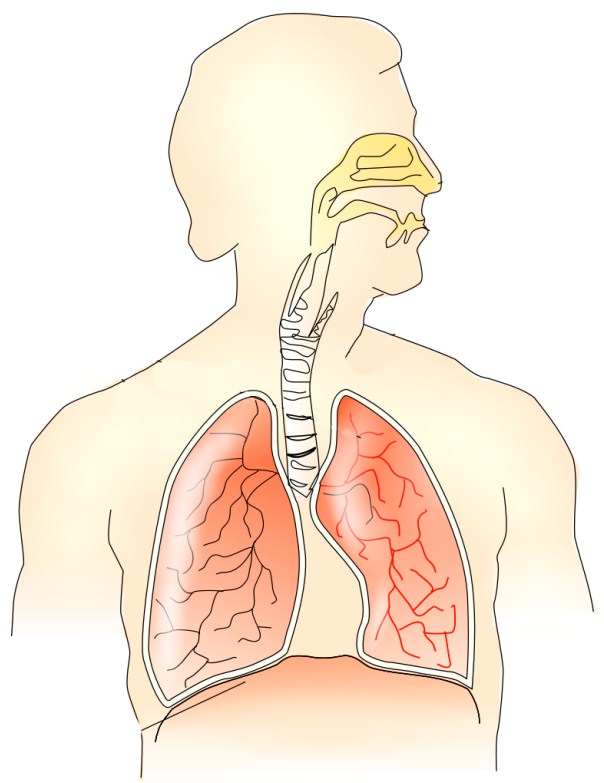
FileRespiratory system complete no labels.svg
The respiratory system. The process of physiological respiration includes two major parts: external respiration and internal respiration. External respiration, also known as breathing, involves both bringing air into the lungs (inhalation) and releasing air to the atmosphere (exhalation). During internal respiration, oxygen and carbon dioxide.
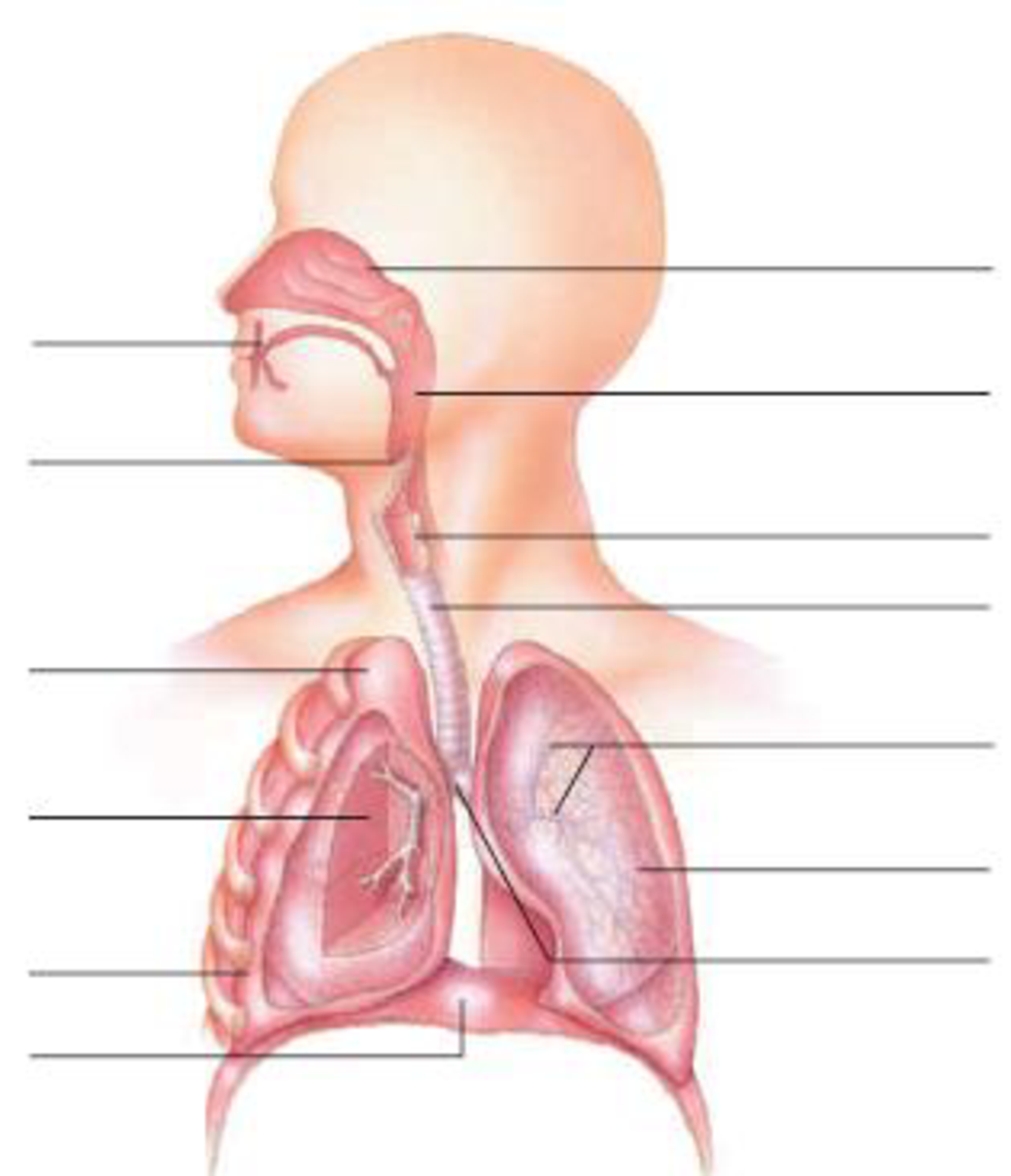
In the diagram below, label the parts of the respiratory system and the structures that enclose
Respiratory system (Systema respiratorum) The respiratory system, also called the pulmonary system, consists of several organs that function as a whole to oxygenate the body through the process of respiration (breathing).This process involves inhaling air and conducting it to the lungs where gas exchange occurs, in which oxygen is extracted from the air, and carbon dioxide expelled from the body.

respiratory system without labels
The organs of the respiratory system form a continuous system of passages called the respiratory tract, through which air flows into and out of the body. The respiratory tract has two major divisions: the upper respiratory tract and the lower respiratory tract. The organs in each division are shown in Figure 16.2.2 16.2.

Respiratory System Diagram to Label Best Of Respiratory System Diagram Unlabeled Respiratory
The major organs in the respiratory system are labeled. [Return to Figure 11.1]. Figure 11.2 image description: This figure shows a cross section view of the nose and throat. The major parts are labeled. [Return to Figure 11.2]. Figure 11.3 image description: This figure shows a micrograph of pseudostratified epithelium. [Return to Figure 11.3].

[DIAGRAM] Diagram Of The Lungs With Labels
You can use the following text: For usage in print - copy and paste the line below: "Respiratory system diagram - no labels" at AnatomyTOOL.org by Mariana Ruiz Villarreal (Wikimedia user: LadyofHats) is in the Public Domain. For digital usage (e.g. in PowerPoint, Impress, Word, Writer) - copy and paste the line below (optionally add the icon):
Respiratory System Diagram Blank Images & Pictures Becuo
This chart of the RESPIRATORY SYSTEM shows how you breathe. Breathing is the process that brings oxygen in the air into your lungs and moves oxygen and through your body. Our lungs remove the oxygen and pass it through our bloodstream, where it's carried off to the tissues and organs that allow us to walk, talk, and move.Our lungs also take carbon dioxide from our blood and release it into the.

THE RESPIRATORY SYSTEM DiveHub.id
2. Select a reference photo. Choose a reference photo online or from your textbook to follow along with. 3. Draw the frame for the diagram. Outline the thoracic cavity, neck, and head. You will want this to be large enough for you to draw more detailed structures inside. For a small diagram, outline the thoracic cavity to be no smaller than 3.