
Mrs. Conner's Science Place Microscope Parts
3. Phase Contrast Microscope. Phase Contrast Microscope is an optical microscope that converts small phase shifts in light into differences in light intensity developing more contrast in images that can be easily detected by human eyes. When light passes through transparent specimens a small phase shift occurs which can't be detected by our eyes.

Microscope Diagram Labeled, Unlabeled and Blank Parts of a Microscope
Free Shipping Available. Buy Part Microscope on ebay. Money Back Guarantee!
13 Tips You Should Know about Taking Care of Your Microscope Rs' Science
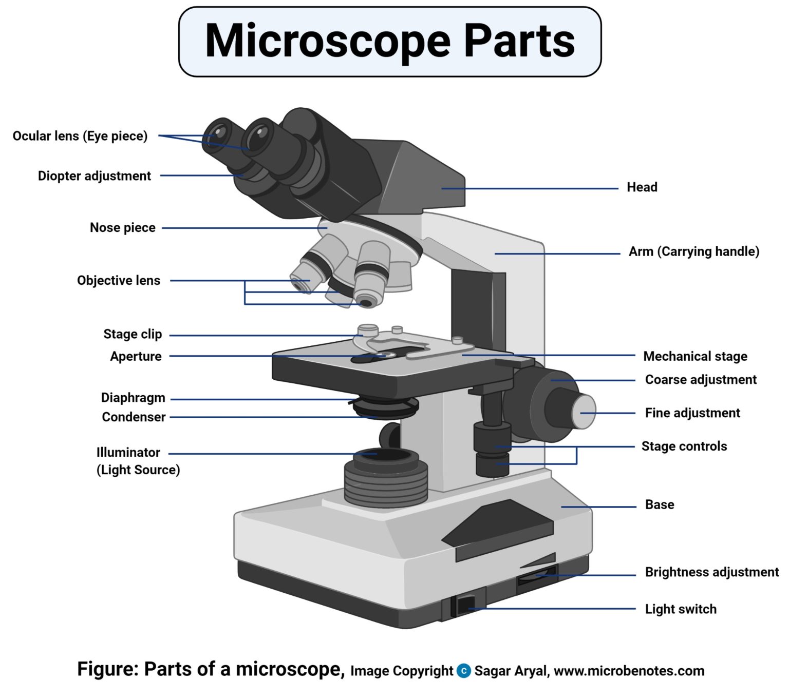
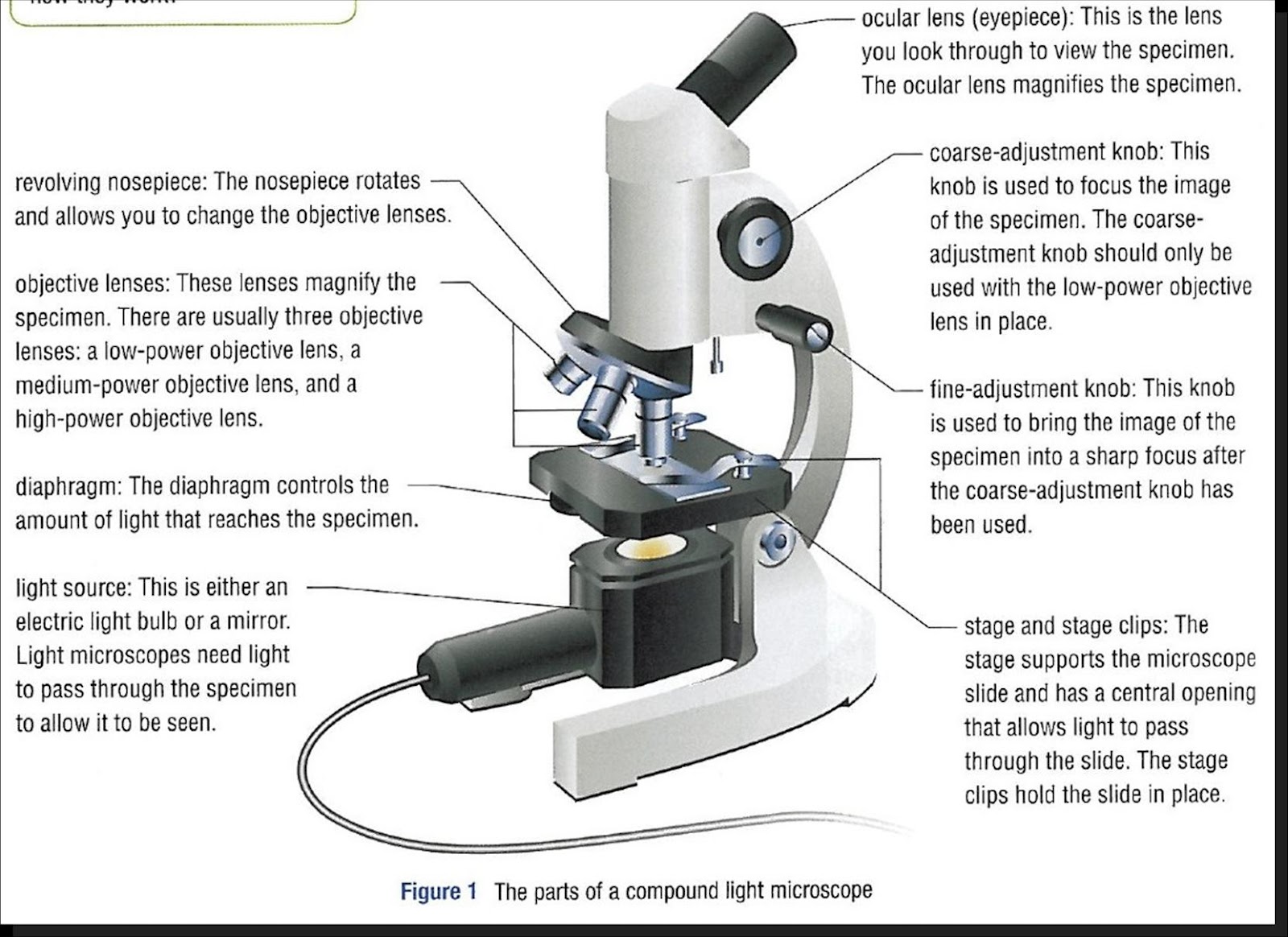
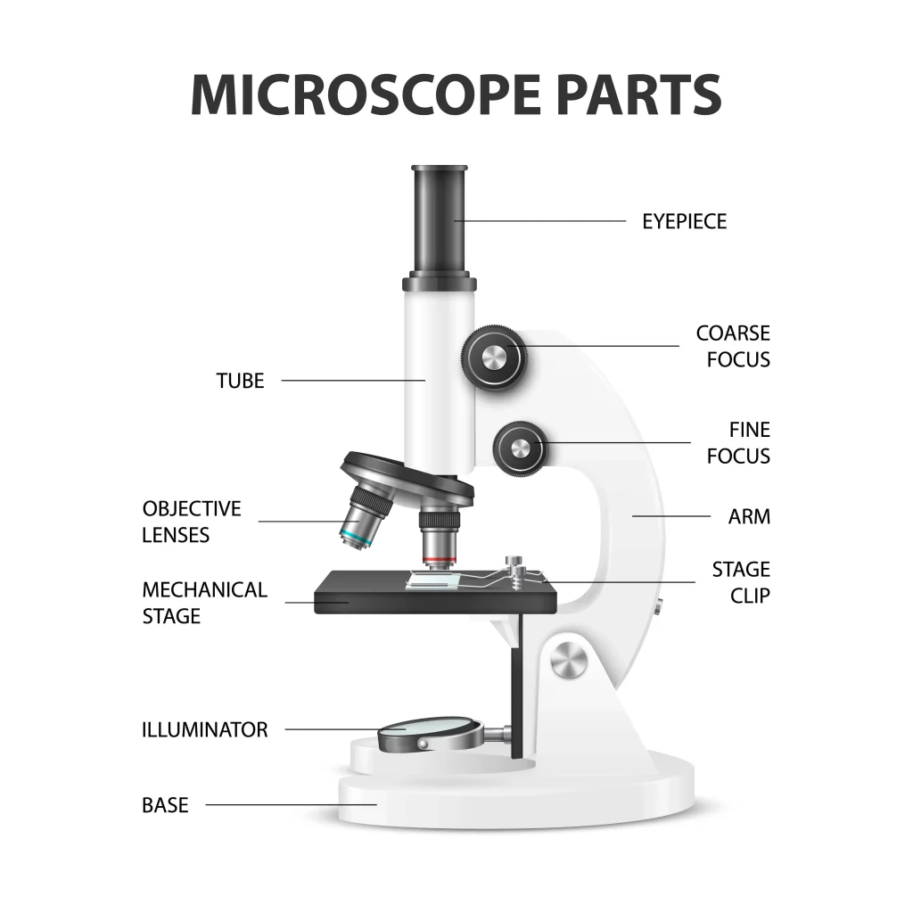
Ocular Lens (eye-piece) Ocular lens of a microscope. It is located at the top of the microscope, and the ocular lens or eyepiece lens is used to look through the specimen. It also magnifies the image formed by the objective lens, usually ten times (10x) or 15 times (15x). Usually, a microscope has an eyepiece of 10x magnification power.
Microscope World Blog Biological Microscope Parts
Microscope Parts & Specifications. Historians credit the invention of the compound microscope to the Dutch spectacle maker, Zacharias Janssen, around the year 1590 (more history here).The compound microscope uses lenses and light to enlarge the image and is also called an optical or light microscope (versus an electron microscope).
SCB 115 Lab 2 Microscope and pH, Acids, Bases, and Buffers Natural Sciences Open Educational
Knobs. Knobs are an important part of any microscope with multiple lenses. They allow the user to do a few different things. The main one is to manually adjust the height of the microscope and move the objective lens closer or farther away from the specimen being magnified. This is known as the coaxial, fine adjustment, or fine focus knob.

Microscope Diagram Labeled, Unlabeled and Blank Parts of a Microscope Tim's Printables
Browse 1,788 microscope parts photos and images available, or search for scanning electron microscope to find more great photos and pictures. Browse Getty Images' premium collection of high-quality, authentic Microscope Parts stock photos, royalty-free images, and pictures. Microscope Parts stock photos are available in a variety of sizes and.
15 Microscope Parts A Guide on their Location and Function
Labomed 9135010 CxL Binocular Cordless Microscope, 4x, 10x, 40x Objectives, LED Illumination. $741.00. ACCU-SCOPE EXM-150-MS Monocular Cordless Microscope with Mechanical Stage, Rechargeable. $351.90. Get relevant offers, the latest promotions, and articles from New York Microscope Company. Parts of a Compound Microscope Each part of the .
Light Microscope Main Parts Of Light Microscope Biology —
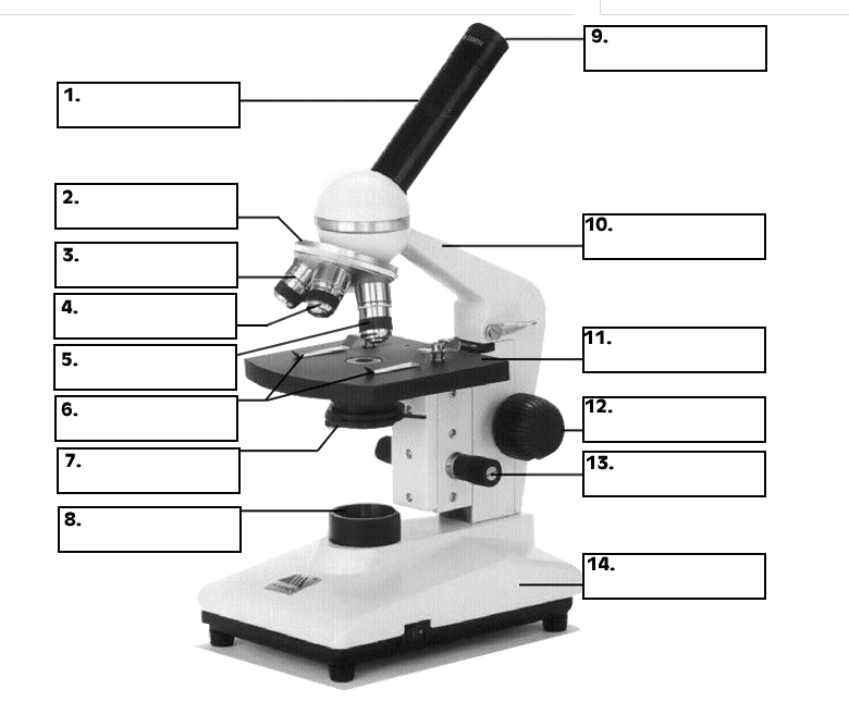
Labeling the Parts of the Microscope. This activity has been designed for use in homes and schools. Each microscope layout (both blank and the version with answers) are available as PDF downloads. You can view a more in-depth review of each part of the microscope here.

Compound Microscope Diagram (Parts labelled), Principle and Uses
Parts of Simple Microscope (Labeled Pictures) It is a magnifying glass with a double convex lens and has a distinct short focal length. It magnifies the object being studied through angular magnification. Below are the vital and complete parts of a simple microscope. Eyepiece/Ocular.
Parts Parts And Functions Of A Microscope
All microscopes share features in common. In this interactive, you can label the different parts of a microscope. Use this with the Microscope parts activity to help students identify and label the main parts of a microscope and then describe their functions.. Drag and drop the text labels onto the microscope diagram. If you want to redo an answer, click on the box and the answer will go back.

36 Label Parts Of The Microscope Labels 2021
5. Knobs (fine and coarse) By adjusting the knob, you can adjust the focus of the microscope. The majority of the microscope models today have the knobs mounted on the same part of the device. Image 5: The circled parts of the microscope are the fine and coarse adjustment knobs. Picture Source: bp.blogspot.com.
What Are The Parts Of A Tree And Their Functions Design Talk
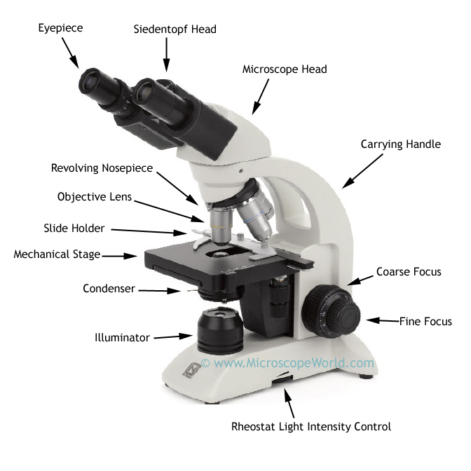
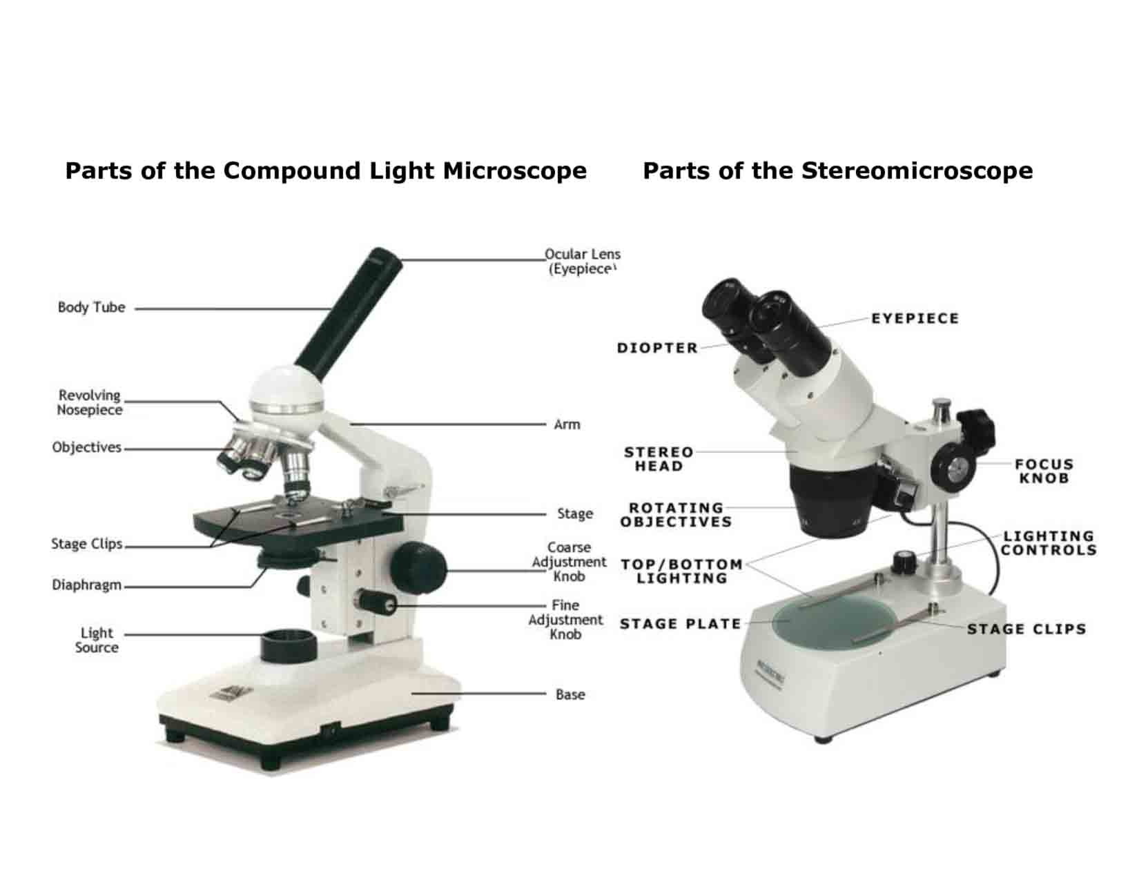
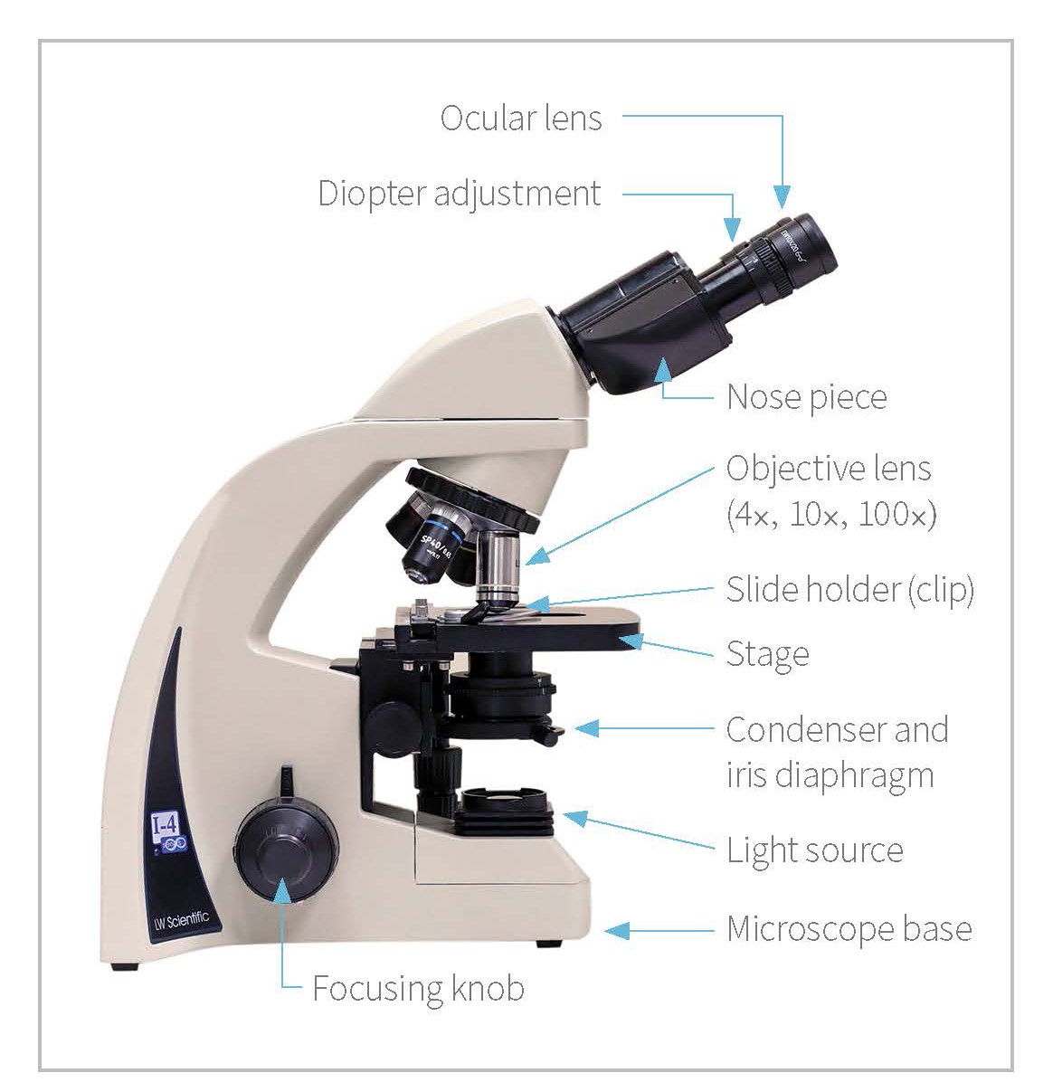
Eyepiece: The lens the viewer looks through to see the specimen. The eyepiece usually contains a 10X or 15X power lens. Diopter Adjustment: Useful as a means to change focus on one eyepiece so as to correct for any difference in vision between your two eyes. Body tube (Head): The body tube connects the eyepiece to the objective lenses. Arm: The arm connects the body tube to the base of the.
Microscopes 7th Grade Science
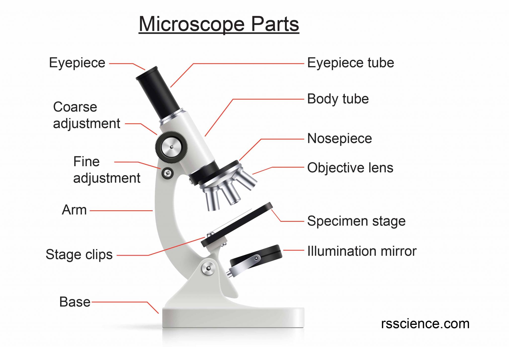
Figure: Diagram of parts of a microscope. There are three structural parts of the microscope i.e. head, arm, and base. Head - The head is a cylindrical metallic tube that holds the eyepiece lens at one end and connects to the nose piece at other end. It is also called a body tube or eyepiece tube.
Parts of a microscope with labeled diagram and functions Biology Notes Web
Microscope Description. A microscope is a laboratory instrument used to examine objects that are too small to be seen by the naked eye. In other words, it enlarges images of small objects. Invented by a Dutch spectacle maker in the late 16th century, light microscopes use lenses and light to magnify images.
Proper Use & Care of Microscopes Veterinary Team Brief
microscope, instrument that produces enlarged images of small objects, allowing the observer an exceedingly close view of minute structures at a scale convenient for examination and analysis.Although optical microscopes are the subject of this article, an image may also be enlarged by many other wave forms, including acoustic, X-ray, or electron beam, and be received by direct or digital.

🎉 Main components of a light microscope. Parts of a microscope with functions and labeled
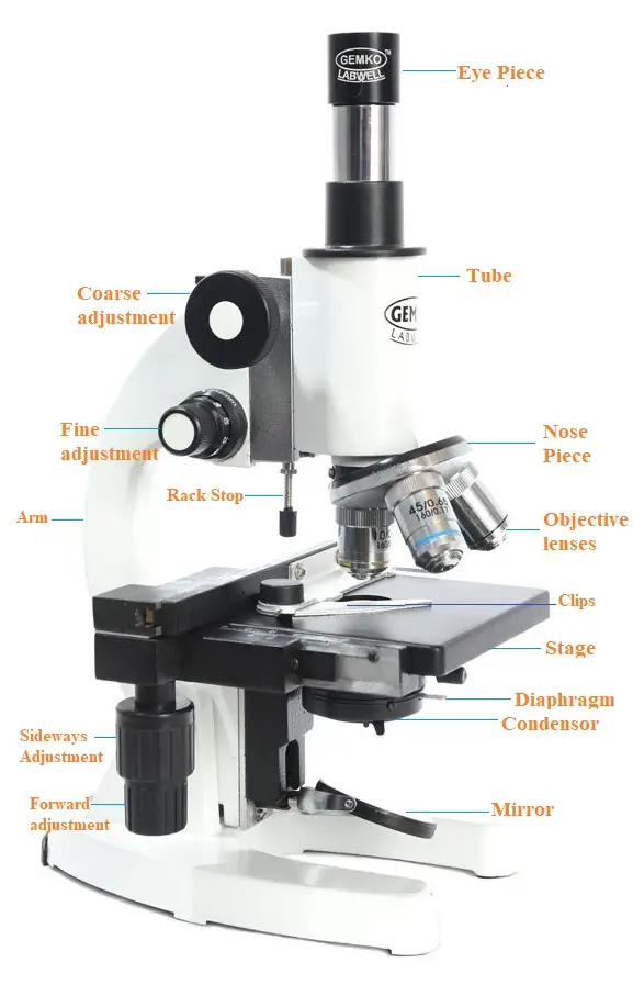
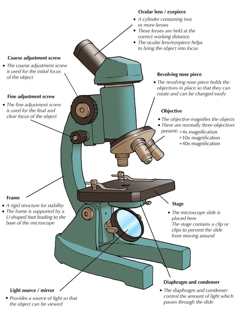
Parts of the optical parts are as follows: Mirror - A simple microscope has a plano-convex mirror and its primary function is to focus the surrounding light on the object being examined. Lens - The biconvex lens is placed above the stage and its function is to magnify the size of the object being examined.