
draw cilia/flagella 9+2 arrangement Brainly.in
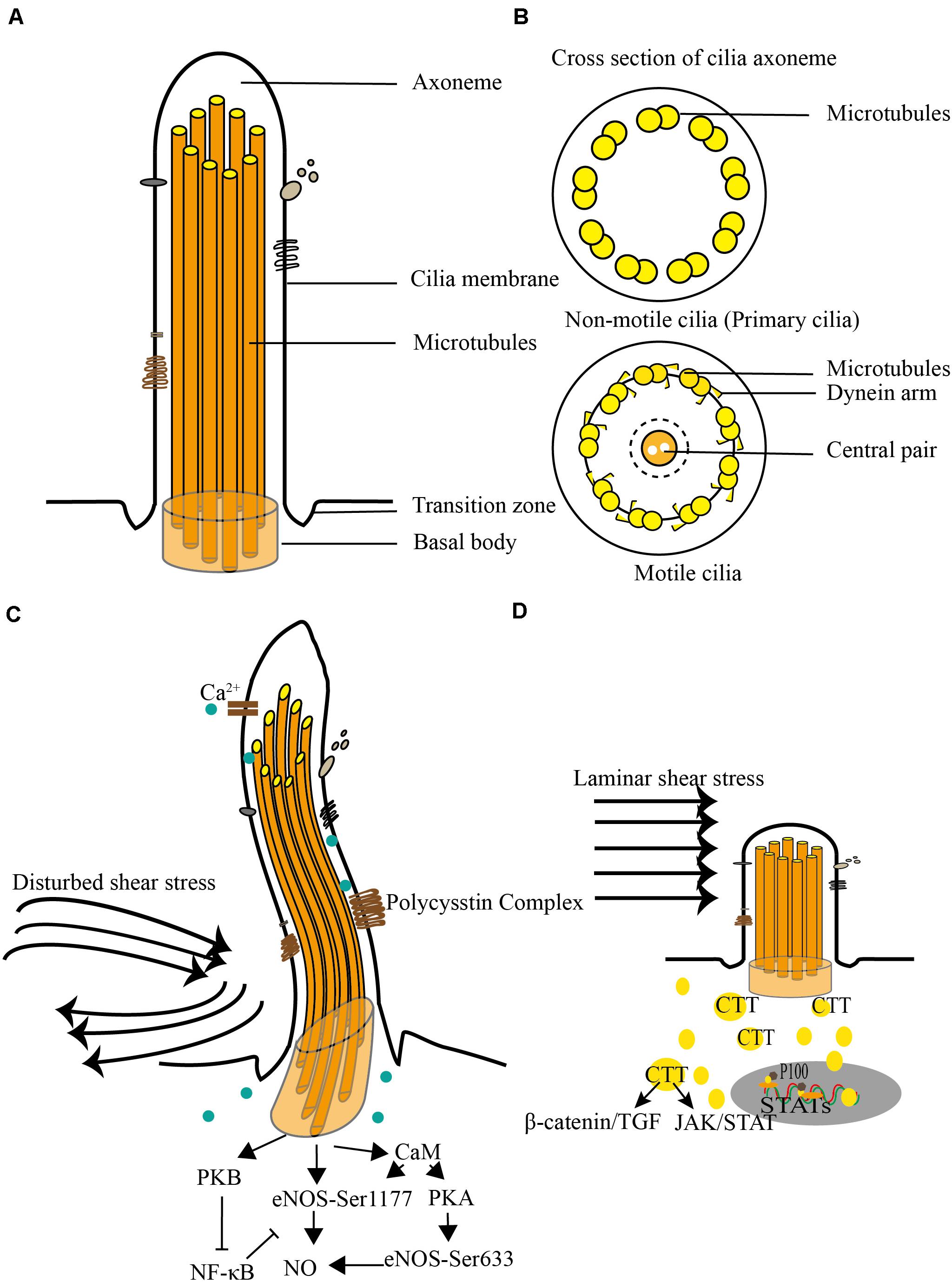
All cilia and flagella are constructed using the same basic framework: The axoneme is a bundle of microtubules that is surrounded by a membrane that is a component of the plasma membrane and is 1 to 2 nm in length and 0.2 m in diameter.
.PNG)
Cell Types and Cell Structure Presentation Biology
Cilia (L. cilium =eye lash) and flagella (Gr. flagellum - whip) are fine hair-like protoplasmic outgrowths of cells and take part in cell motility. These organelles were first reported by Englemann (1868). Cilia and flagella are basically similar but they vary in number, length and patterns of movement.
Which Of The Following Is Not A Function Cilia And Flagella About Flag Collections
Cilia and flagella are formed from specialized groupings of microtubules called basal bodies. If the protrusions are short and numerous they are termed cilia. If they are longer and less numerous (usually only one or two) they are termed flagella. What Are Their Distinguishing Characteristics?

Cilia and flagella biological structure difference comparison outline diagram. Labeled
Flagella (singular = flagellum) are long, hair-like structures that extend from the plasma membrane and are used to move an entire cell, (for example, sperm, Euglena ). When present, the cell has just one flagellum or a few flagella. When cilia (singular = cilium) are present, however, they are many in number and extend along the entire surface.
Animal Cell Definition, Structure, Parts, Functions, Labeled Diagram
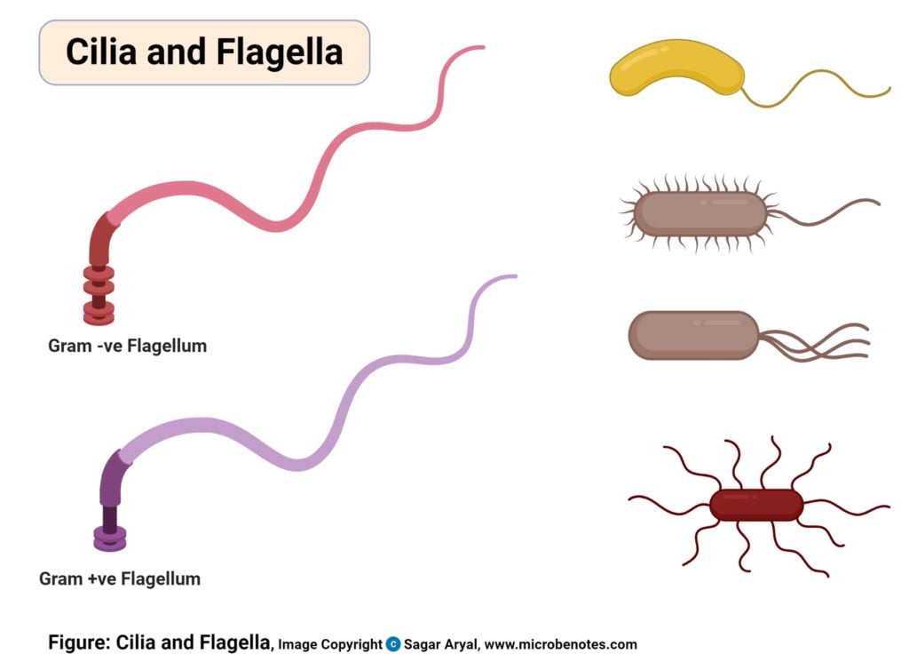
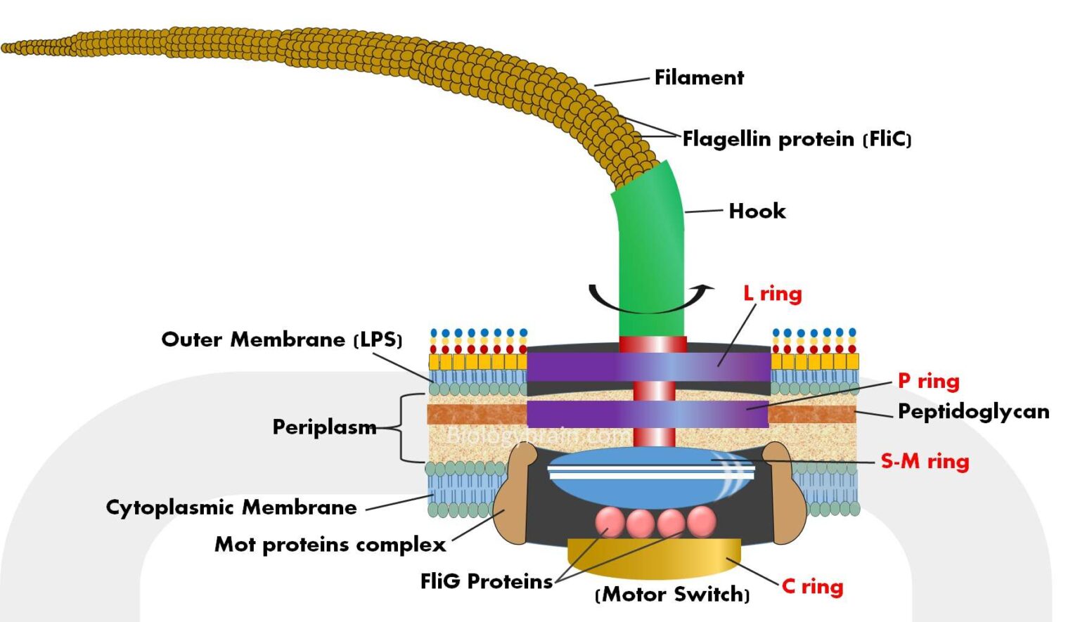
Cilia and Flagella Frequently Asked Questions on Flagella Bacterial Flagella Structure The flagella is a helical structure composed of flagellin protein. The flagella structure is divided into three parts: Basal body Hook Filament Basal Body It is attached to the cell membrane and cytoplasmic membrane.

Flagellum & Cilia Wikimedia Commons CCA 3.0 Unported by Urutsegh and Kohidai Cell Structure
So this right over here is a picture of the amoeba Chaos carolinense. And what you see here is a projection coming off from the main part of the cell, and this is called a pseudopod, which is referring to it being a false foot. The pod is coming from the same root word as podiatry, which is referring to the foot.
Flagellum microscope hires stock photography and images Alamy
5.6: Flagella and Cilia. Flagella (singular = flagellum) are long, hair-like structures that extend from the plasma membrane and are used to move an entire cell, (for example, sperm, Euglena ). When present, the cell has just one flagellum or a few flagella. Prokaryotes sometimes have flagella, but they are structurally very different from.

Blink Activity BlinkLearning
A flagellum or flagella is a lash or hair-like structure present on the cell body that is important for different physiological functions of the cell. The term 'flagellum' is the Latin term for whip indicating the long slender structure of the flagellum that resembles a whip.

Schematic drawing of eukaryotic flagella ultrastructure (A)... Download Scientific Diagram
Structure of a cell > Tour of a eukaryotic cell The cytoskeleton The cytoskeleton. Microtubules, microfilaments (actin filaments), and intermediate filaments. Centrioles, centrosomes, flagella and cilia. Introduction What would happen if someone snuck in during the night and stole your skeleton?

2 rings in the basal body Google Search Plasma membrane, Microbiology, Cell wall
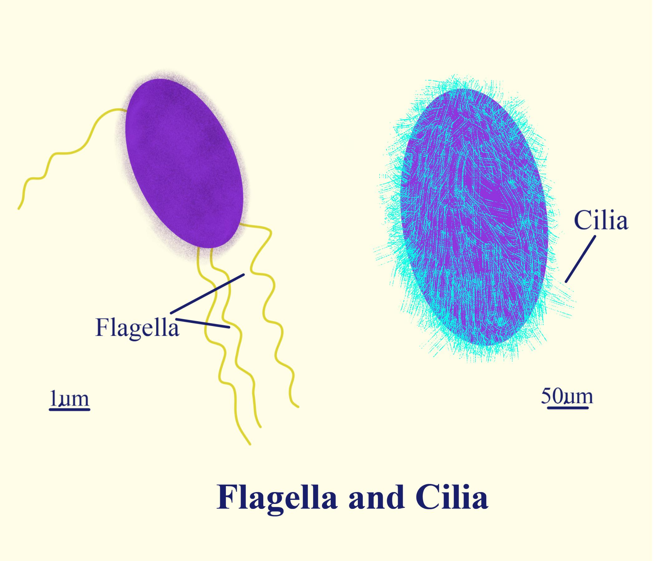
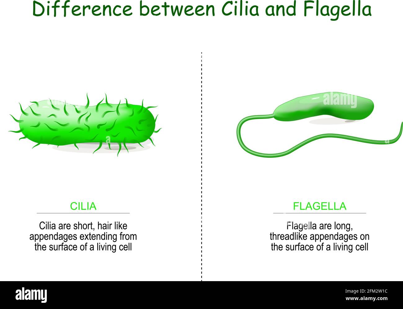
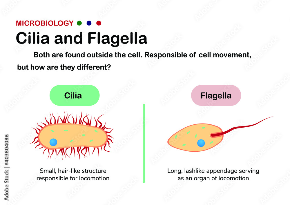
Cilia and flagella are cell organelles that are structurally similar but are differentiated based on their function and/or length. Cilia are short and there are usually many (hundreds) cilia per cell. On the other hand, flagella are longer and there are fewer flagella per cell (usually one to eight).

Top 115+ Cilia and flagella plant or animal cell
Cilia and flagella are fine, whiplike/hairlike structures that extend from the body of a variety of cells. While they vary in terms of length and numbers in different types of cells (as well as patterns of movement), cilia and flagella are generally identical in structure and composition.

Difference Between Cilia And Flagella In Eukaryotes cloudshareinfo
Cilia and flagella have the same internal structure. The major difference is in their length. Cilia and flagella move because of the interactions of a set of microtubules inside. Collectively, these are called an "axoneme", This figure shows a microtubule (top panel) in surface view and in cross section (lower left hand panel)..
Frontiers Primary Cilia and Atherosclerosis Physiology
Structure of Flagella and Cilia: They are fine hair like movable protoplasmic processes of the cells which are capable of producing a current in the fluid medium for locomotion and passage of substances. Flagella are longer (100-200 µm) but fewer. Only 1-4 flagella occur per cell, e.g., many protists, motile algae, spermatozoa of animals.
Flagella Definition Structure Types Arrangement Functions Examples Riset
Nature Education 3 (9) :54 What is a primary cilium? Learn how an organelle can be both a sensing organ and a transport machine. Aa Aa Aa Eukaryotic flagella and cilia have long been recognized.
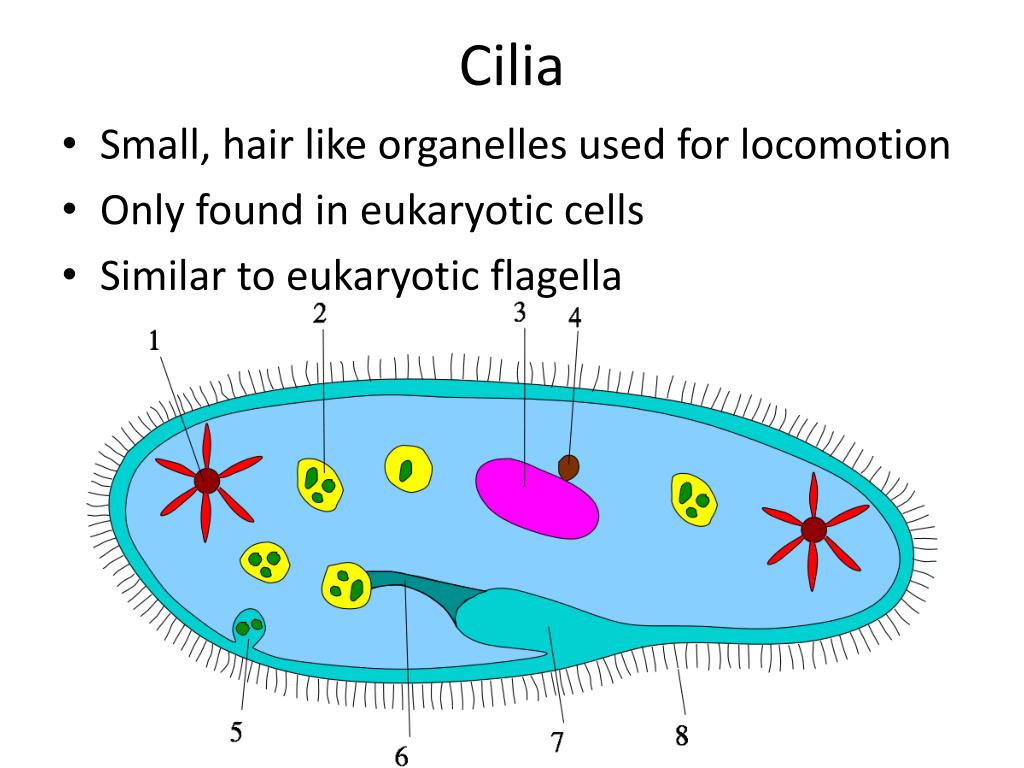
PPT Cilia and Flagella in Cell Structure PowerPoint Presentation, free download ID333456
The bending of cilia (and flagella) has many parallels to the contraction of skeletal muscle fibers. Testing the Model. Remember, the partial microtubules do not extend as far into the tip as the complete microtubules. So if a slice is made a short distance back from the tip: A straight cilium should show the complete pattern (center of diagram).
Biology diagram present different of cilia and flagella in eukaryote and prokaryote organism
Figure 7.7.7 7.7. 7 .7.3: A cilium (plural cilia) is an organelle found in eukaryotic cells. Cilia are slender protuberances typically extending some 5-10 micrometers outwards from the cell body. There are two types of cilia: motile cilia, which constantly beat directionally, and non-motile—or primary—cilia, which typically serve as.