Beyond the obvious Exploring Os Tibiale Externum and Os Peroneum in Foot and Ankle Pain A
Accessory navicular. The accessory navicular is also known as the os tibiale, os tibiale externum, or naviculare secundarium. Estimated prevalence has been set in between 4 and 21% [1,2,3].A recent long series by Kalbouneh et al. estimates it as 20.9% [].The accessory navicular is located adjacent to the postero-medial tuberosity of the bone, and three different configurations exist [].
Os Tibiale Externum Ortobas
The os tibiale externum(1) also called the accesory navicular is the most commonly found accesory ossicle of the foot with reported incidence of about 25-30%. It is located on the posteromedial aspect of the foot adjacent to the posteromedial tuberosity of the navicular bone. Three types of accessory tibiale externum have been described in.

Lower Extremity Os Foot & Ankle Orthobullets
Also known as 'os tibiale externum' or 'os navicularum', accessory navicular syndrome refers to a congenital abnormality related to the growth of an extra bone within the foot. This additional piece of bone is not present in a normal human foot and grows toward the middle inner part of the foot near the navicular bone.
Knickfuss Leonardo
An accessory bone or supernumerary bone is a bone that is not normally present in the body, but can be found as a variant in a significant number of people. It poses a risk of being misdiagnosed as bone fractures on radiography. [2] Wrist and hand [ edit] X-ray of the wrist, with most common accessory bones labeled. [3]

Os tibiale externum sagittal T2 YouTube
The accessory navicular—also known as the os naviculare or os tibiale externum—is a small bone that extends from the navicular bone, one of the tarsal bones near the instep. About 14 percent of the population has an accessory navicular, and about half of the people with the extra bone have it in both feet.
Os tibiale externum DocCheck
The accessory navicular (os navicularum or os tibiale externum) is an extra bone or piece of cartilage located on the inner side of the foot just above the arch. It is incorporated within the posterior tibial tendon, which attaches in this area and can lead to Accessory Navicular Syndrome. An accessory navicular is congenital (present at birth).

Os tibiale externum Image
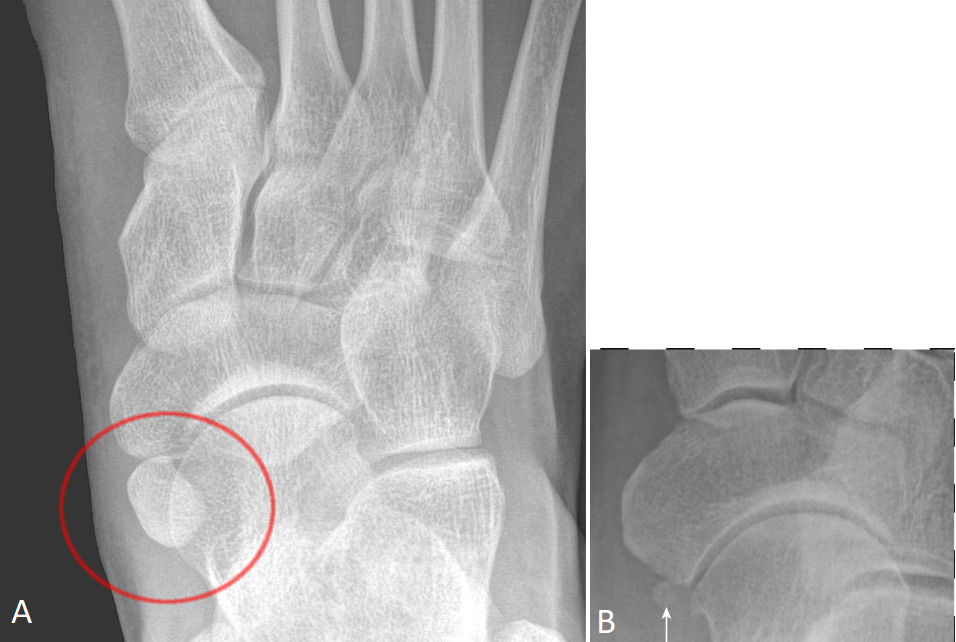
Classification This classification was proposed by Geist 7 in 1914 and remains the most widely used classification system (c. 2021). The Geist classification divides these into three types: type 1 accessory navicular bone (os tibiale externum, os naviculare secundarium)

Os tibiale externum Image
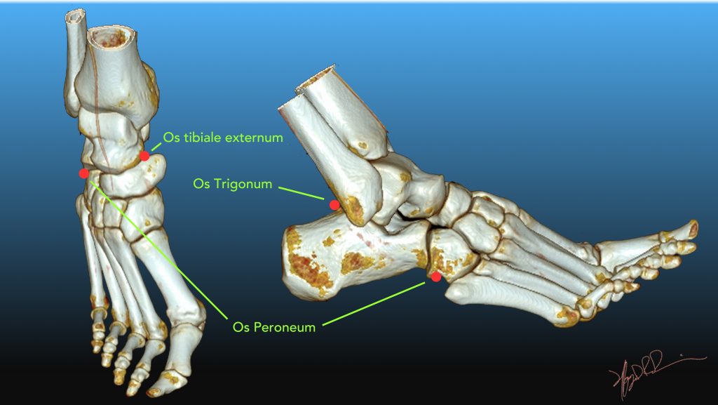
Etiology Definition accessory ossicles secondary ossification centers that remain separated from the normal bon sesamoids are bones that are incorporated into tendons and move with normal and abnormal tendon motion Most common ossicles os trigonum accessory navicular (os tibiale externum) os intermetatarseum Most common sesamoids os peroneum

Os_tibiale_externum Don't the Bubbles
os tibiale externum (accessory navicular) os trigonum os calcaneus secundaris os calcanei accessorium 6 os intermetatarseum pars peronea metatarsalis primi (pars peronea metatarsalia) os supratalare bipartite hallux sesamoid os supranaviculare os infranaviculare (cuneonavicular ossicle) 5 os intercuneiforme os vesalianum pedis os sustentaculi

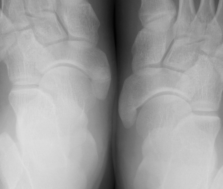
Os tibiale externum type II Image
The accessory navicular (os navicularum or os tibiale externum) is an extra bone or piece of cartilage located on the inner side of the foot just above the arch. It is incorporated within the posterior tibial tendon, which attaches in this area. An accessory navicular is congenital (present at birth).
Common Accessory Ossicles of the Foot UW Emergency Radiology
The accessory navicular syndrome, also known as os naviculare syndrome occurs when a type II accessory navicular becomes painful due to movement across the pseudo-joint between the ossicle and the navicular bone.. Radiographic features Ultrasound. It can be inferred on musculoskeletal ultrasound if a patient's pain is located at a type II accessory navicular and the patient is tender to.

Lower Extremity Os Foot & Ankle Orthobullets
The os tibiale externum is also known as accessory navicular bone, os naviculare secundarium, accessory (tarsal) scaphoid, or prehallux. It is found within the tibialis posterior tendon near its insertion on the navicular bone. The os peroneum is a small sesamoid bone located within the peroneus longus tendon, adjacent to the cuboid.
Os Tibiale Externum Ortobas
Accessory navicular syndrome (ANS) happens when an abnormal foot growth called the navicular bone (os navicularum or os tibiale externum) creates pain on the side of the foot between the medial arch and heel.

Zusätzliches Kahnbein (Os naviculare accessorium, Os tibiale externum ) fussInfo
Type I is a 2-3 mm sized sesamoid bone, also referred to as os tibiale externum and is located at the level of the inferior calcaneonavicular ligament within the tibialis posterior tendon. Type II is an accessory bone, also referred to as prehallux , connected to the navicular by a fibrocartilage or hyaline cartilage (synchondrosis).

Os tibiale externum Image
Os tibiale externum (OTE) also termed accessory navicular, os naviculare, or os navicularis is a common accessory bone in the foot located medial and sometimes proximal to the navicular tuberosity. It is attached and continuous with the tibialis posterior tendon and is present in 10 to 15% of the population either unilateral or bilateral.
ostibialeexternumtypeiiandiii KENSHIN blog
Os tibiale externum (accessory navicular) is a large ossicle adjacent to the medial side of the navicular bone. The tibialis posterior tendon often inserts with a broad attachment onto the ossicle, which may cause a painful tendinosis due traction between the ossicle and the navicular. Such changes are best seen on MRI. Credit: Dr Donna D'Souza.