Mega cisterna magna Kinderneurologie
Introduction Fetal central nervous system (CNS) abnormalities are fairly common, with an incidence of about 0.1%-0.2% in live births and an even higher occurrence of about 3%-6% in stillbirths. They are second only to cardiac malformations in terms of frequency of occurrence ( 1 ).

18 Mega Cisterna Magna Radiology Key
The resilient North American auto industry, one of the most powerful engines driving the economy in this region, continues to be shaped by globalization and the power of innovation. New technologies and systems are accelerating the pace of change in an industry that is poised to provide transportation solutions for the 21st century.

Mega cisterna magna Radiology Case
What is Dandy-Walker syndrome? Dandy-Walker syndrome is sometimes called Dandy-Walker malformation or just Dandy-Walker. It's a congenital brain malformation that causes an issue with how the brain forms. It is congenital meaning a baby is born with the condition, and it occurs as the baby develops during pregnancy.

Mega cisterna magna Image
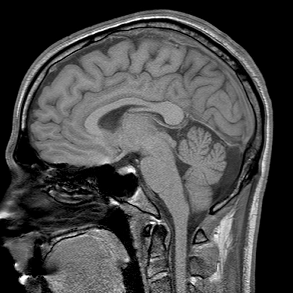
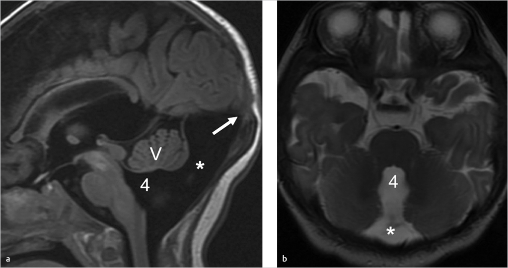
Cisterna Magna. A mega cisterna magna (also called a retrocerebellar arachnoid pouch, communicating arachnoid cyst, and Blake pouch) is a developmental variation of the posterior fossa characterized by an enlarged bony/dural posterior fossa, expansion of the cisterna magna (which freely communicates with the perimedullary subarachnoid spaces), and an intact vermis with a normal fourth.

Mega cisterna magna Image
Mega cisterna magna refers to a cystic posterior fossa malformation that is characterized by an enlarged cisterna magna, absence of hydrocephalus, and an intact cerebellar vermis. It must be differentiated from persistent Blake's pouch cyst, posterior fossa cysts, Dandy-Walker variant, and posterior fossa neoplasia.

Mega cisterna magna Image
The cisterna magna is a cerebrospinal fluid (CSF) filled space located in the posterior fossa dorsal to the medulla and caudal to the cerebellum. Mega cisterna magna refers to a cystic posterior fossa malformation characterized by an enlarged cisterna magna, absence of hydrocephalus, and an intact cerebellar vermis. [1] [2]

Mega cisterna magna Radiology Case
The authors present two cases of mega cisterna magna associated with mania and catatonic schizophrenia. Dandy-Walker malformation is a rare congenital abnormality characterized by large posterior fossa cyst with open communication between the fourth ventricle,.

Mega Cisterna Magna StatPearls NCBI Bookshelf
The cisterna magna is located between the cerebellum and the dorsal surface of the medulla oblongata at and above the level of the foramen magnum. CSF produced in the ventricular system drains into the cisterna magna from the fourth ventricle via the median aperture (of Magendie) and the lateral apertures (of Luschka) 1,2.

Mega Cisterna Magna on MRI Stock Image M210/0398 Science Photo Library
Results: Subjects with isolated mega cisterna magna had a lower performance on memory tasks [RAVLT saving score (0.8 +/- 0.2 vs. 1.02 +/- 0.2, P = 0.003)] and verbal fluency [phonemic fluency (9.4 +/- 4.5 vs. 13.6 +/- 5.3, P = 0.02), semantic fluency (19.8 +/- 5.8 vs. 24.4 +/- 7.5, P = 0.05)].

Mega cisterna magna Radiology Case
Mega Cisterna Magna (MCM) MCM refers to a retro- and infracerebellar CSF space greater than 10 mm on mid-sagittal images. It is thought to arise during embryogenesis from a defect of the posterior membranous area of the roof of the fourth ventricle, with mildly deficient fenestration of Blake's pouch ( Robinson and Ederies, 2016 ).

Mega Cisterna Magna Prominent cisterna magna appearing as a wedge shaped area of CSF density
A mega cisterna magna is thought to occur in ~1% of all brains imaged postnatally. It constitutes 54% of all cystic posterior fossa malformations 4. Associations Especially if noted antenatally, a mega cisterna magna has been associated with: infarction inflammation/infection: particularly cytomegalovirus

Mega cisterna magna Radiology Case
Cisterna magna is a normal CSF (cerebrospinal fluid) filled space on the backside of the brain. However, in many individuals, it can be more extensive, called mega cisterna magna, and that usually does not cause any symptom to the patient. It needs to be differentiated from an arachnoid cyst or cerebellar atrophy, or dandy walker syndrome.
Mega Cisterna Magna
A mega cisterna magna is a controversial subject. In general, however, the term refers to non-pathological enlargement of the retrocerebellar CSF space, not associated with cerebellar abnormalities (normal cerebellar vermis and hemispheres).
18 Mega Cisterna Magna Radiology Key
Hordeaux J, Hinderer C, Goode T, et al. Toxicology study of intra-cisterna magna adeno-associated virus 9 expressing human alpha-L-iduronidase in rhesus macaques. Mol Ther Methods Clin Dev. 2018;10:79-88.

Mega cisterna magna Radiology Case
A mega-cisterna magna, is a controversial entity among experts. In general however, the term is applied to non-pathological prominance (usually exceeding 10 mm in antenatal imaging) of the retro-cerebellar CSF space and not associated with cerebellar abnormalities. There is a normal vermis and normal cerebellar hemispheres.

Mega cisterna magna Radiology Case
What are mega cisterna magna (MCM) and arachnoid cysts (AC)? MCM involves the enlargement of normal fluid-filled space in the brain with no other structural differences anomaly in other cerebral structures. ACs are usually benign, cerebro-spinal fluid-like collections that develop within the layers of the membranes that wrap the brain.