Flexor retinaculum Physiopedia
The hamulus also serves as the attachment point for a number of different muscles and ligaments of the hand and forearm, including the flexor retinaculum. Articulations The hamate bone articulates with several adjacent bones: The proximal surface articulates with the lunate bone;

Flexor Retinaculum of Hand Anatomy l Surface marking l Structures Passing l Superficial l Deep
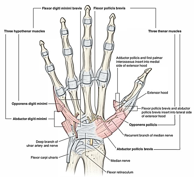
The Palmar carpal ligament (PCL) is a distinct component of the antebrachial fascia. The distal part and the true covering of the carpal tunnel is the flexor retinaculum. There is an area in the distal part of the flexor retinaculum that consists of crisscrossing of muscle aponeurosis of the thenar and hypothenar muscles ( Fig. 18.9a,b ).
Flexor Retinaculum (Hand) Earth's Lab
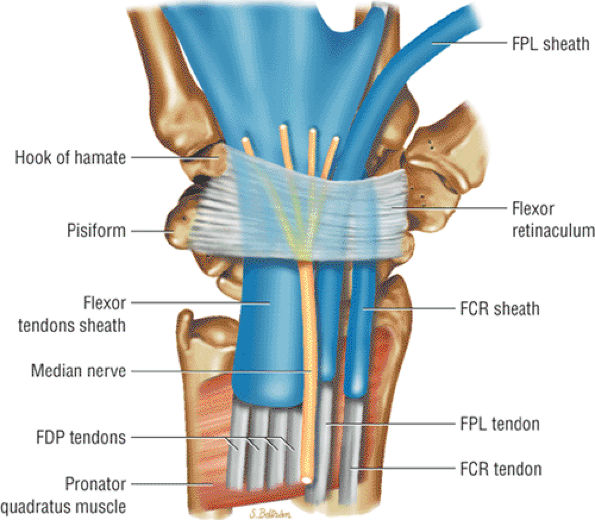
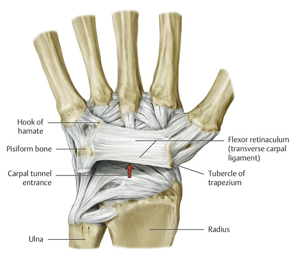
Flexor Retinaculum Thick connective tissue which forms the roof of the carpal tunnel. Turns the carpal arch into the carpal tunnel by bridging the space between the medial and lateral parts of the arch. Spans between the hook of hamate and pisiform (medially) to the scaphoid and trapezium (laterally).

15 The Forearm Fascia and Retinacula Musculoskeletal Key
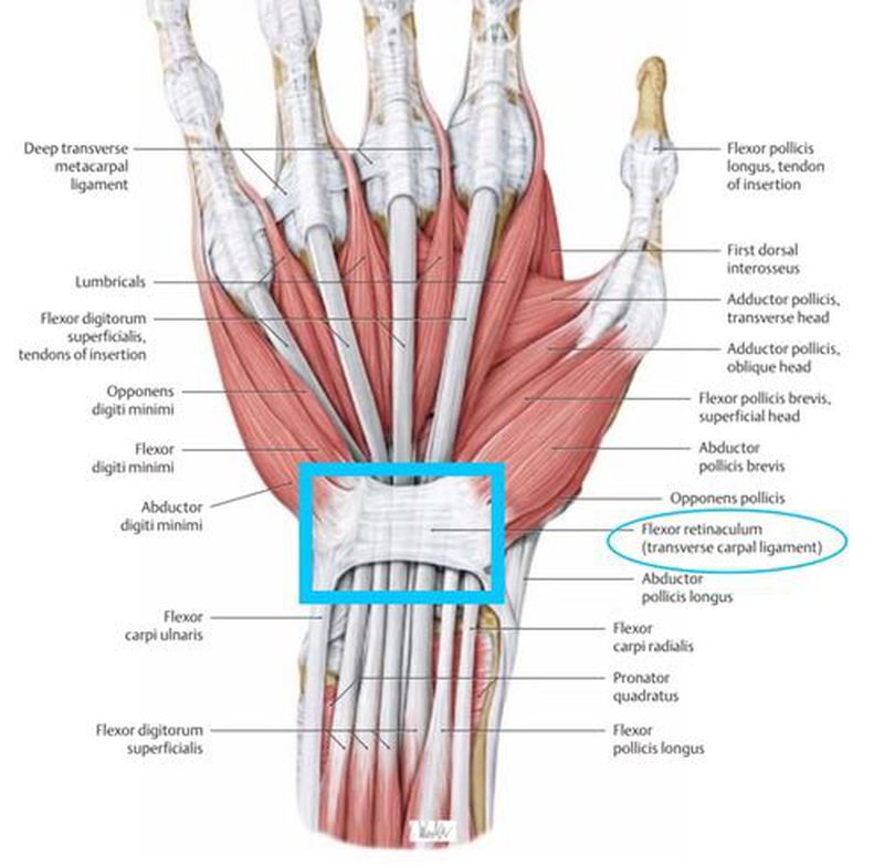
The flexor retinaculum ( transverse carpal ligament, or anterior annular ligament) is a fibrous band on the palmar side of the hand near the wrist. It arches over the carpal bones of the hands, covering them and forming the carpal tunnel . Structure
The Wrist and Hand TeachMe Orthopedics
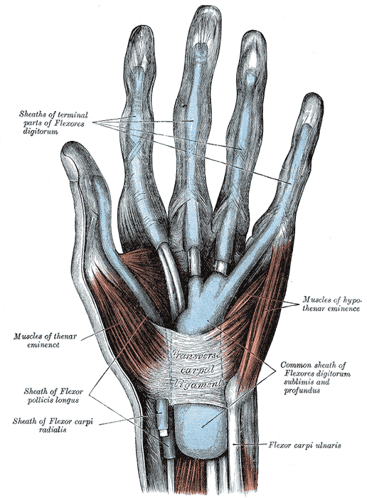
Definition The flexor retinaculum (transverse carpal ligament; anterior annular ligament) is a strong, fibrous band, which arches over the carpus, converting the deep groove on the front of the carpal bones into a tunnel, through which the Flexor tendons of the digits and the median nerve pass.

Superior Extensor Retinaculum Anatomy, Musculoskeletal system, Orthopedic surgery
Retinaculum. A retinaculum refers to any region on the body in which tendon groups from different muscles pass under one connective tissue band. Wrist retinacula include the flexor and the.
The Forearm, Wrist, and Hand Musculoskeletal Key
1/4 Synonyms: none Intercarpal joints are all classified as synovial plane joints, meaning that the articular surfaces are functionally considered as nearly flat and lined with fibrocartilage. The joints are enclosed by the thin fibrous capsules whose internal surfaces are lined by the synovial membranes.
Flexor Retinaculum (Hand) Earth's Lab
Flexor Tendon Injuries are traumatic injuries to the flexor digitorum superficialis and flexor digitorum profundus tendons that can be caused by laceration or trauma.. Zone is unique in that FDP and FDS in same tendon sheath (both can be injured within the flexor retinaculum). Tendons can retract if vincula are disrupted.
Flexor retinaculum (Retinaculum flexorum) Kenhub
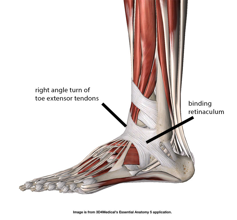
The flexor retinaculum of the foot is a strong fibrous band that covers the tendons of the muscles that flex the foot such as walking on the toes like a ballerina.

The flexor retinaculum in the carpal tunnel consists of three segments... Download Scientific
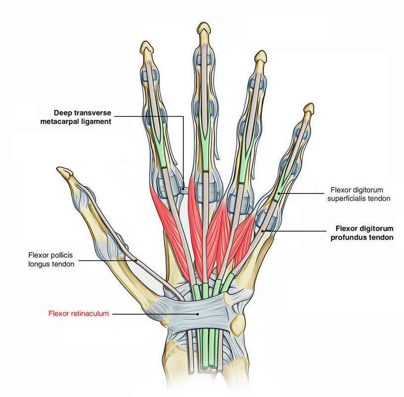
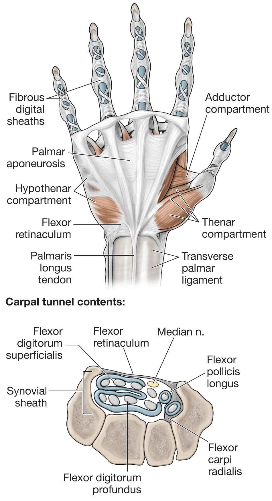
The roof of the carpal tunnel is formed by the flexor retinaculum (also known as transverse carpal ligament), a thick connective tissue ligament. This ligament bridges the space between the medial and lateral ends of the carpal arch, converting the arch into a tunnel. Contents Tendons of flexor digitorum profundus muscle
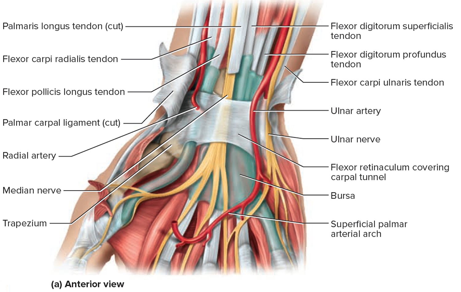
View of the wrist showing the flexor retinaculum at the wrist and the
The flexor retinaculum (also known as the transverse carpal ligament ) is a rectangular-shaped fibrous band located at the volar aspect of the hand, near the wrist. Gross anatomy The flexor retinaculum encloses and forms the roof of the carpal tunnel. The ulna aspect of the flexor retinaculum forms the floor of Guyon's canal.
The Mechanical Function of Retinacula Academy of Clinical Massage
The flexor tendon sheaths of the remaining three fingers are separate. The radial bursa extends for the entire length of the flexor pollicis longus tendon and ends just proximal to the flexor retinaculum. The radial & ulnar bursa communicate at the level of the wrist joint in almost 50% of individuals. Dorsal carpal tendinous sheaths
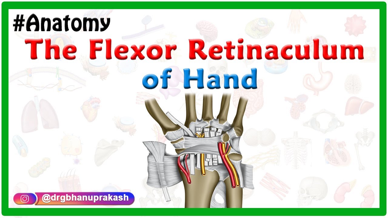
The flexor retinaculum of Hand Gross anatomy , Attachments and Relations YouTube
The flexor retinaculum is a fibrous connective tissue band that forms the anterior roof of the carpal tunnel (see Image. Flexor Retinaculum of the Wrist). Many experts consider the flexor retinaculum synonymous with the transverse carpal and annular ligaments.

Pin em Musculoskeletal System
Flexor retinaculum is a strong fibrous band which bridges the anterior concavity of the carpal bones thus converts it into a tunnel, the carpal tunnel [1]. Attachments Medially, To the pisiform bone To the hook of the hamate Laterally, To the tubercle of the scaphoid To the crest of the trapezium [1]
Anatomy and Functional Anatomy of the Hand Plastic Surgery Key
Definition The TCL is the middle portion of the flexor retinaculum (FR). 1 The proximal portion of the FR is the distal continuation of the antebrachial fascia. 2 The transition from the antebrachial fascia to the TCL can be identified based on gross inspection, predominantly marked by the abrupt increase in thickness.
Flexor Retinaculum MEDizzy
The flexor retinaculum at the ankle is formed by reinforcement of the deep fascia of the leg by transverse collagen bundles and functions to prevent 'bowstringing' of tendons as they pass the tibiotalar joint. It forms the roof of the tarsal tunnel 1-2. Attachments medial malleolus of the tibia medial process of the calcaneus plantar aponeurosis