
Structure of interosseous talocalcaneal ligament Semantic Scholar
5 Interosseous talocalcaneal ligament 6 Cervical talocalcaneal ligament Akiyama suggested that the sinus tarsi is not only a talocalcaneal joint space but a source of nociceptive and proprioceptive information on the movement of the foot and ankle and that sinus tarsi syndrome may result from disorders of nociception and proprioception in the foot.

Interosseous Talocalcaneal ligament Nursing School Info, Nursing Study, Body Health, Health
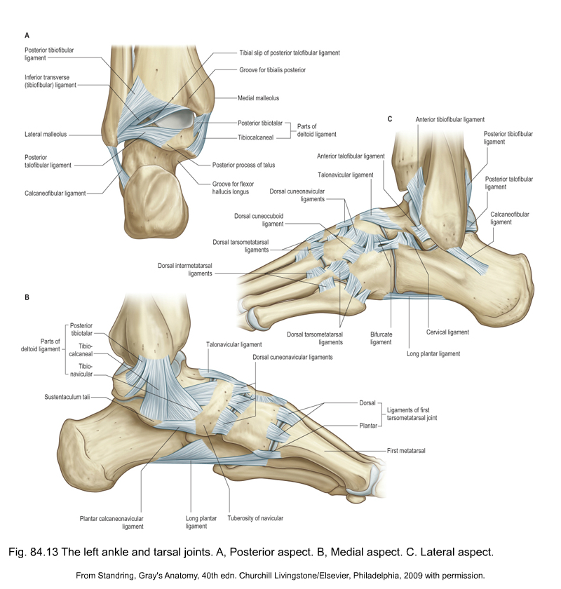
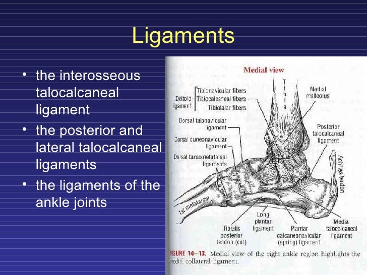
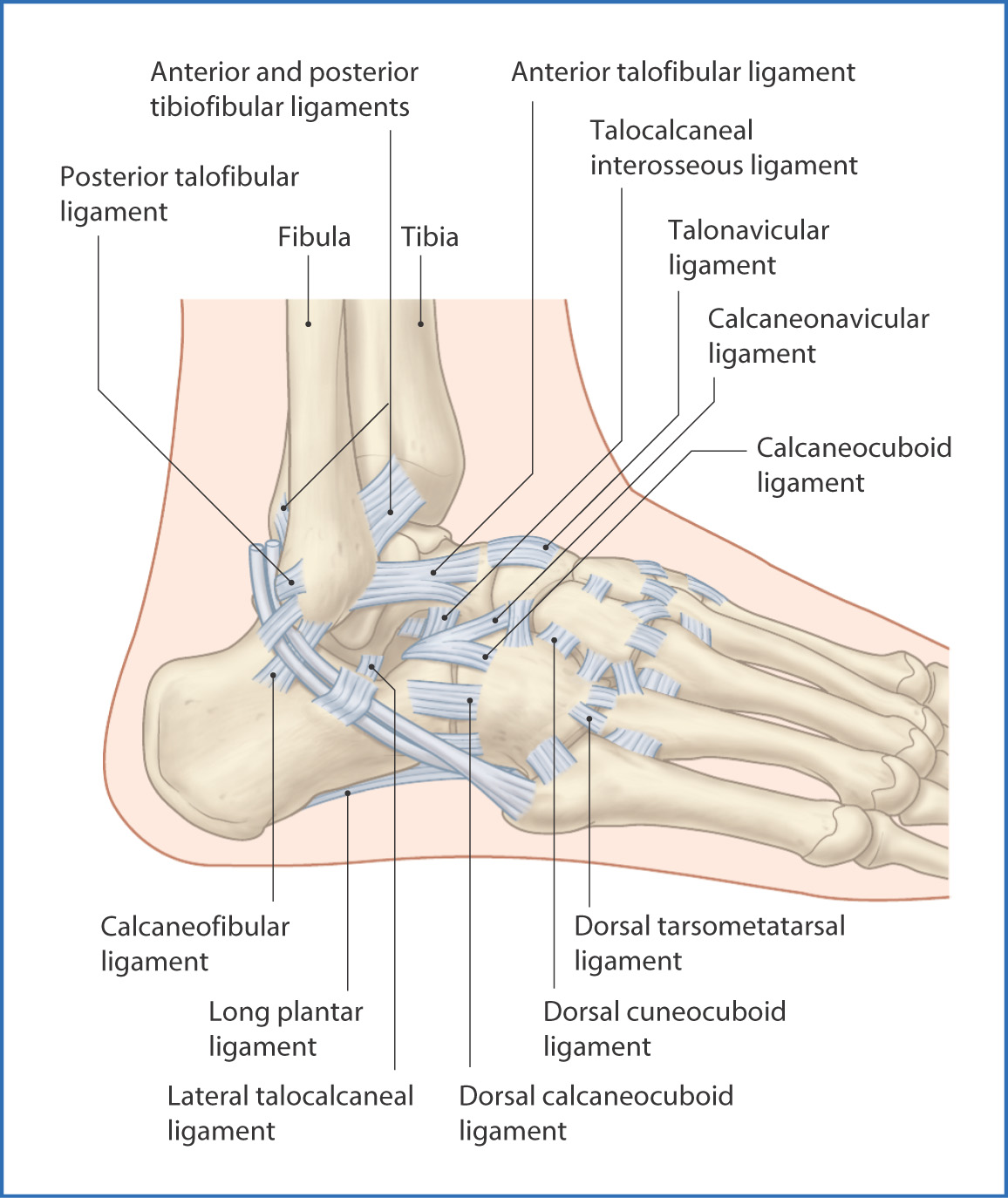
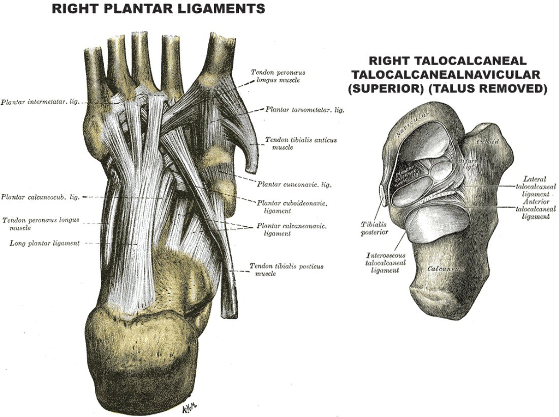
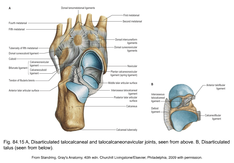
The interosseous talocalcaneal ligament is located in the sinus tarsi and is broad, flat and bilaminar. The posterior lamina of the ligament is associated with the talocalcaneal joint, and anterior lamina with the talocalcaneonavicular joint. Its medial fibers are taut in eversion. References
anatomy of the lower leg, ankle and foot Musculoskeletal Key
Background: Injury of the interosseous talocalcaneal ligament (ITCL) has been recognized as a cause of subtalar instability, though lack of an accepted clinical test has limited the ability of clinicians to reliably make the diagnosis. Clinical effects of ITCL failure remain unclear because of insufficient understanding of the role of the ligament.

The interosseous talocalcaneal ligament. a Posterior view specimen with... Download Scientific
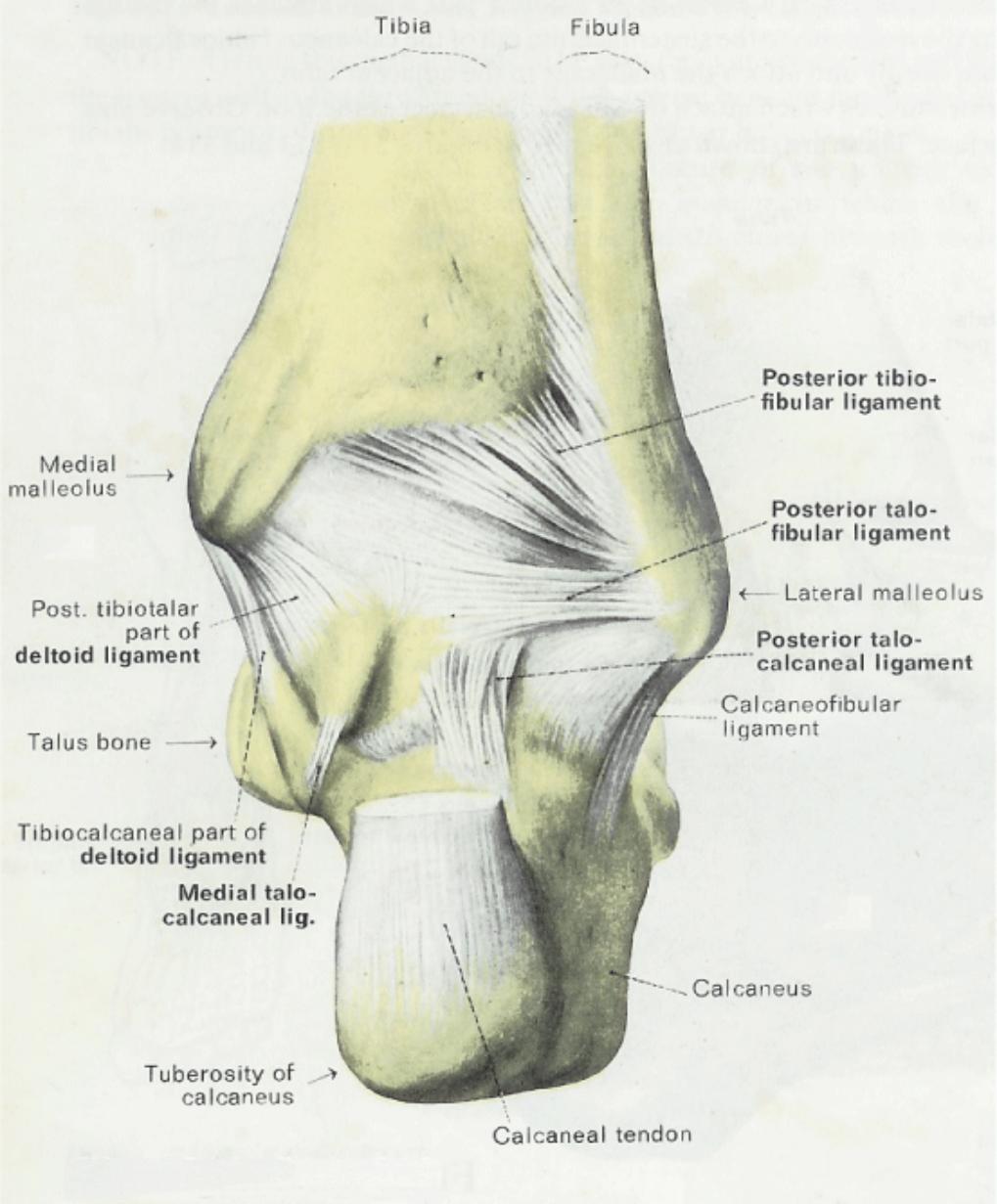
The interosseous talocalcaneal ligament forms the chief bond of union between the bones. It is, in fact, a portion of the united capsules of the talocalcaneonavicular and the talocalcaneal joints, and consists of two partially united layers of fibers, one belonging to the former and the other to the latter joint. It is attached,above, to the groove between the articular facets of the under.

(A) T1weighted MRI scan with a coronal view showing rupture of... Download Scientific Diagram
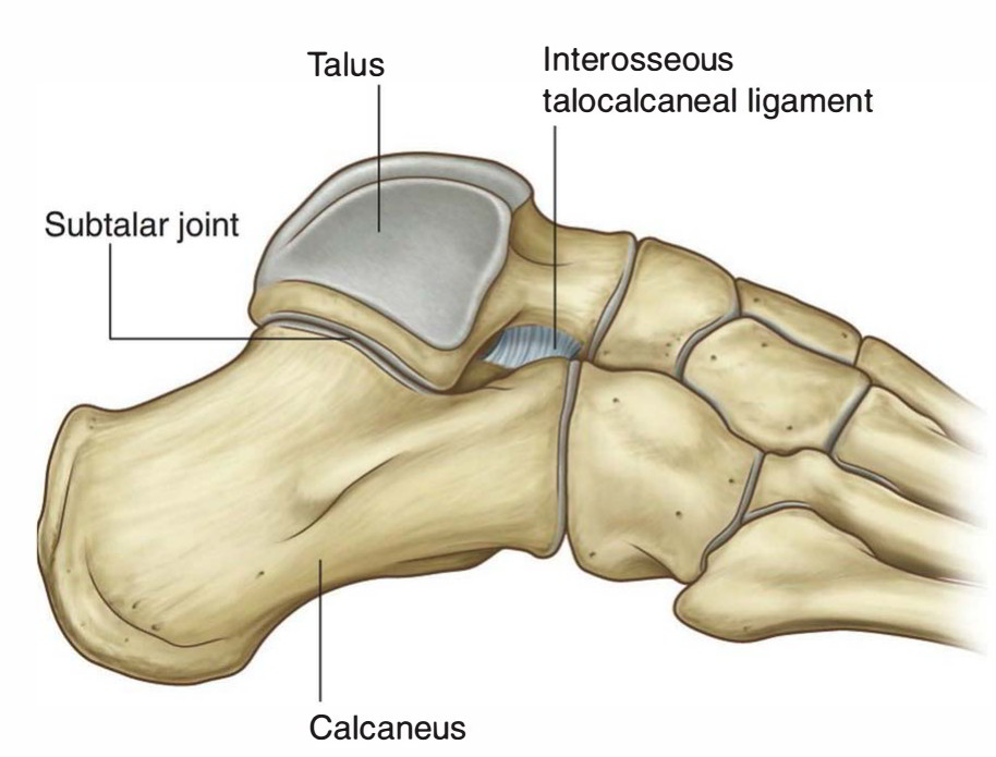
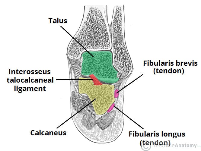
The talocalcaneal interosseous ligament is the main stabilizer of the subtalar joint. It occupies a central position between the subtalar and talocalcaneonavicular joints and lies directly below the long axis of the leg, thus acting as the fulcrum around which the leg and foot moves. This subjects the talocalcaneal interosseous ligament to.

Anatomy Of The Ankle
Lateral talocalcaneal ligament An additional ligament - the interosseous talocalcaneal ligament - acts to bind the talus and calcaneus together. It lies within the sinus tarsi (a small cavity between the talus and calcaneus), and is particularly strong; providing the majority of the ligamentous stability to the joint.
Kinesiology Of Ankle And Foot
The anterior talocalcaneal ligament ( anterior calcaneo-astragaloid ligament or anterior interosseous ligament) is a ligament in the foot . The anterior talocalcaneal ligament extends from the front and lateral surface of the neck of the talus to the sinus tarsi of the calcaneus . It forms the posterior boundary of the talocalcaneonavicular joint .
Anatomy & Physiology Illustration
The interosseous talocalcaneal ligament (ITCL) is the main soft-tissue contributor to subtalar joint stability. The role of ITCL reconstruction in retaining this stability is minimally reported. Therefore, we conducted this study to investigate the effects of rupture and reconstruction of the ITCL on the subtalar and peritalar joints.
Talocalcaneal Ligament Earth's Lab
The interosseous talocalcaneal ligament forms the chief bond of union between the talus and calcaneus . It is a portion of the united capsules of the talocalcaneonavicular and the talocalcaneal joints, and consists of two partially united layers of fibers, one belonging to the former and the other to the latter joint.
Ankle and Foot Joints Basicmedical Key
This part (also known as the "true" joint capsule) forms the strong talocalcaneal interosseous ligament, together with the anterior part of the talocalcaneal joint capsule. The joint capsule is lined with the synovial membrane which helps to lubricate the joint to facilitate movements of the bones.

The posterior talocalcaneal ligaments connections in two different... Download Scientific Diagram
The main ligament that attaches these bones is called the interosseous talocalcaneal ligament, which runs along a groove between them. Four other weaker ligaments provide the joint with added stability. In between the calcaneus and talus is the synovial membrane. This tissue secretes fluid to lubricate the joint space, protecting the cartilage.
Anatomy Of The Right Foot
The interosseous talocalcaneal ligament is composed of two short and broad fibrous bands located in the tarsal sinus. The deep extension of the inferior extensor retinaculum is situated between these bands of the interosseous ligament. Occupying the central position between the talocalcaneal and talocalcaneonavicular joints, this ligament is.
Interosseous talocalcaneal ligament. (a) Schematic drawing of a coronal... Download Scientific
Its stability resides in the configuration of the anterior and posterior subtalar joint surfaces as well as several ligamentous structures (Fig. 1) [6, 25, 27]: the inferior extensor retinaculum (IER), the calcaneo-fibular ligament (CFL), the cervical ligament (CL) and finally the inter-talocalcaneal ligament (ITCL), which comprises an anterior and a posterior fascicle that rotate over each.

The interosseous talocalcaneal ligament. a Posterior view specimen with... Download Scientific
The main stabilizing ligaments of the subtalar joint (STJ) are the cervical ligament (CL) and the interosseous talocalcaneal ligament (ITCL). The primary function of the CL is to resist excessive STJ supination whereas the ITCL remains taut during pronation. Dysfunction of either the CL or ITCL can result in subtalar instability, resulting in a.
The Subtalar Joint Ligaments Neurovascular TeachMeAnatomy
The interosseous talocalcaneal ligament and the medial component of the extensor retinaculum root form a V-shape in the tarsal sinus and canal. Function. The talocalcaneal interosseous ligament controls the talus in the movements of eversion and inversion by maintaining apposition of the talus and calcaneus. The function of the cervical.
anatomy of the lower leg, ankle and foot Musculoskeletal Key
Subtalar joint instability is an entity commonly neglected within the scope of lateral ankle instability in patients of all ages; up to 25% of chronic ankle instabilities have associated subtalar instability [2, 24, 30], which could account for some of the cases with persistent symptoms after isolated repair or reconstruction of the anterior talo-fibular ligament [].